IVF

Performing ICSI using inverted microscope and special microcontrollers.

ICSI is applied when the sperm is incapable of fertilising the egg on its own (as previously described). Using highly specialised equipment, the embryologist deposits a single spermatozoon inside the egg to induce activation and fertilisation of the egg. Only one motile spermatozoon is required for each egg.

The method has been successfully used since 1992 and overcomes almost all the barriers that cause male infertility.

Thanks to ICSI, men with severely compromised sperm quality (reduced number, low motility, bad morphology or ejaculation problems) can now father a child of their own; something that was impossible in the not so distant past.

Until today, thousands of babies have been born from ICSI, with no indication that the method presents a danger for embryo quality and child health (according to a large scale epidemiological study from Belgium).

Except the way that fertilization occurs, the procedure is the same as conventional IVF as far as the man and woman are concerned.

Indications for ICSI

ICSI was developed to treat mainly male infertility. Indications include:

  • Severe oligo-asthenozoospermia
  • Azoozpermia
  • Globozoospermia (absence of acrosome)
  • Sperm agglutination
  • P revious fertilization failure with conventional IVF in the presence of normal sperm
  • ICSI can also be used for other reasons, such as low number or poor quality of oocytes, think or hard zona pelucida, especially in older women. However, this approach is in doubt by many scientists.

Oocyte with normal fertilization with 2 pronuclei and 2 polar bodies.

The following morning after oocyte retrieval, i.e. 16-20 hours after insemination the fertilization check is performed. The embryologists check under the microscope how many oocytes have been fertilized normally and isolate unfertilized or abnormally fertilized oocytes. Normal fertilization is verified by the presence of 2 pronuclei (2PN) and 2 polar bodies.

Schematic representation of the fertilization process.

The chromosomes of the spermatozoon and the oocyte have been condensed and are enveloped by a membrane, giving rise to the two pronuclei (1 male and 1 female). At the same time the second polar body is extruded marking the completion of meiosis II. The first cells of the new embryo is now called zygote and has the same size and shape as the oocyte.

A proportion of the inseminated oocytes after IVF or ICSI either do not form pronuclei, or form only I pronucleus (1PN) or multiple pronuclei (3PN, 4PN). After conventional IVF, the presence of multiple PNs means that the oocyte was fertilized by more than just one spermatozoa. These embryos are called polyspermic and have surplus numbers of chromosomes. Polyspermic embryos should not be transferred as they are related in abnormal pregnancies (miscarriages, myli pregnancy).

Usually, normal fertilization rates in the laboratory range from 50% to 100%. It depends on:

  • The quality and maturity of the oocytes
  • The fertilizing ability of the sperm
  • Optimal culture conditions

Polyspermy rates are low (3-5% of oocytes).

IVF, apart from infertility treatment, has also a diagnostic value as it gives an indication that the oocytes and sperm of a couple can interact. In some cases of unexplained infertility we discover a possible cause for infertility and may obtain useful information, such as no attachment of sperm on the zona pelucida, low sperm motility after a few hours of culture etc. The laboratory conditions and culture media are optimized so that they do not affect fertilization in any way.

Fertilization rates do not reflect the quality of resulting embryos and a low fertilization rate should not cause worries about the outcome of IVF.

Embryo development begins a few hours after the formation of the zygote. The zygote divides in two cells that are called blastomeres. The next divisions follow, giving rise to more and smaller blastomeres. This process is called cleavage.

Stages of embryo development until the blastocyst stage

Two days after oocyte retrieval cleavage has started and embryos have undergone 2 cell divisions. Cleavage rates are high, about 95%. Typically, the first division occurs about 16 hours after fertilization (2-cell embryo). The second division happens 12 hours later (4-cell embryo) and divisions continue in an increasing rate. Between 4 and 8-cell stage the embryonic genome is activated. The next stages are the morula and the blastocyst.

Correct cleavage is important for embryo quality. The number and morphology of blastomeres, the presence of fragmentation, the morphology and fusion of pronuclei and the synchronous divisions are important features for the evaluation of embryos and are related to IVF outcome.

Therefore, embryos with late cleavage (e.g. 2-cell stage on day 2) are considered inferior to 4-cell stage embryos.

The development of the zygote up the blastocyst stage

During natural conception, the blastocyst hatches from the embryonic shell, the zona pellucida, on the 5th or 6th day after fertilization. The fully hatched blastocyst is the last free-form embryonic stage and the only stage when the embryo has the capacity to attach and implant in the endometrium. However, in some cases, the zona pellucida is harder or thicker than normal, obstructing the process of hatching and, as a result, impairing successful implantation.

Hatching blastocysts after thawing.
Following an artificial opening on the zone pelloucida using laser the blastocysts have started to hatch.

When the embryos develop in culture, there is another possible intervention before the embryo transfer; the embryologist can assist blastocyst hatching by opening a small hole on the zona pellucida (assisted hatching), using either a special laser device or a chemical solution.

The initial excitement over the usefulness of assisted hatching in implantation has not been widely accepted by embryologists. The method does not seem to significantly increase implantation rates, while it subjects the embryos to further stress. However, assisted hatching has been proven to slightly improve implantation in special cases, such as thick zona, frozen-thawed embryos, eggs from women of increased age, etc.

Eugonia offers the assisted hatching method with the use of laser, in the cases that is has been deemed necessary by our scientific team.

The achievement of a successful pregnancy largely depends on the number and quality of embryos transferred to the uterus. Therefore, we need to evaluate a patient's embryos during their development and select for transfer those with the highest morphological characteristics.

Evaluation of Day 2-3 embryos

Cleavage

On Day 2 after oocyte retrieval the embryos must have 2-4 cells, with optimal stage the 4-cell stage. On Day 3, the embryos must have 5-8 cells, with 8-cell embryos being the best developmentally.

Morphology- Fragmentation

Embryos are categorized into four grades (I-IV) based on size and shape of blastomeres and percentage of fragmentation. Grade I embryos are the best morphologically, without fragmentation, normal size and shape of blastomeres. On the contrary, Grade IV includes poor quality embryos with total fragmentation or degenerated blastomeres. Intermediate Grades are II and III. Ideally, on day 2 we want to have 4-cell embryos with grades I or II. ON day 3, best embryos are regarded 8-cell embryos with grades I or II.

4-cell embryos

4-cell embryos
 

8-cell embryos

8-cell embryos
8-cell embryos

Other criteria

Multinucleation

Normally, each blastocyst of a developing embryo must have one visible nucleus. The presence of multiple nuclei (multinucleation) indicates an abnormal number of chromosomes and is related with a lower chance of implantation.

Uniformity of blastomeres

Uniformity of blastomeres is also related to embryo quality. Normally, a good quality embryo has blastomeres of equal size and shape. However, it is normal for a blastomere to have a larger size than the rest for odd-number cell stage (3, 5,7 cells).

Cytoplasm

The presence of granules or vacuoles inside the blastomeres should be recorded, although these characteristics have not been definitely related to poor embryo quality.

Thickness of zona pelucida

The zona pelucida is an acellular glycoprotein layer that surrounds preimplantation embryos. During expansion of the blastocyst, the zona ruptures and allows the embryo to hatch and implant. Increased thickness of the zona pelucida is related to lower chances of implantation as they embryo may get trapped inside it and fail to hatch successfully.

Evaluation of blastocysts

See: Blastocysts.

Metabolic criteria

See: Metabolomics.


Day 2 embryos

2 cell Grade I
2 cell Grade I
2-cell Grade II
2-cell Grade II
2-cell Grade III
2-cell Grade III
 
3-cell Grade II
3-cell Grade II
 
4-cell Grade I
4-cell Grade I
4-cell Grade II
4-cell Grade II
4-cell Grade II-III
4-cell Grade II-III
4-cell Grade III
4-cell Grade III
4-cell Grade IV
4-cell Grade IV
 

Day 3 embryos

5-cell Grade I
5-cell Grade I
5-cell Grade II
5-cell Grade II
6-cell Grade I
6-cell Grade I
6-cell Grade I-II
6-cell Grade I-II
6-cell Grade II-III
6-cell Grade II-III
6-cell Grade III
6-cell Grade III
6-cell Grade IV
6-cell Grade IV
 
7-cell Grade II
7-cell Grade II
7-cell Grade II-III
7-cell Grade II-III
8-cell Grade I (fertilized with ICSI)
8-cell Grade I  (fertilized with IVF)
8-cell Grade I (fertilized with IVF)
8-cell Grade II
8-cell Grade II
8-cell Grade II-III
8-cell Grade II-III
8-cell Grade III
8-cell Grade III
8-cell Grade IV
8-cell Grade IV
9-cell Grade I
9-cell Grade I
 
12-cell Grade I
12-cell Grade I
 

Day 4 embryos

Morula
Morula
 

Day 5/6 embryos (Blastocysts)

Early blastocyst
Early blastocyst
 
Full blastocyst 3ΑΑ
Full blastocyst 3ΑΑ
Full blastocyst 3ΑΒ
Full blastocyst 3ΑΒ
Full blastocyst ΒΒ
Full blastocyst ΒΒ
Full blastocyst 3CC
Full blastocyst 3CC
Expanded blastocyst 4AA
Expanded blastocyst 4AΒ
Expanded blastocyst 4AC
Expanded blastocyst 4BB
Expanded blastocyst 4BC
Expanded blastocyst 4CC
Hatching blastocyst 5AA
Fully hatched blastocyst 6ΑΑ

Surrogacy applies to couples with normal eggs and sperm but the woman has a non functional uterus or is unfit to gestate for medical reasons.

Fertilization occurs in the embryology laboratory.

The gestation of a couple's embryo by a third woman is allowed by judicial permission according to article 1458 of N.3089/02, provided that there is a documented and unselfish agreement between the couple and the surrogate mother (and her husband if the latter is married).

PGD allows the identifications of genetic abnormalities of the embryo while it develops in culture. The method detects certain genetic abnormalities in the embryo, which are responsible for known congenital or hereditary diseases.

Embryos that undergo PGD are biopsied on Day 3 after oocyte retrieval. The embryos are first placed in a special solution to loosen the intercellular bonds. Then a small opening is created on the zona pelucida using laser, and using a special micropipette one cell (blastomere) is removed from each embryo. The biopsied cell will undergo genetic analysis, while the remaining embryo will continue to develop in culture. It has been observed that the removal of a single blastomere at this stage of embryonic development does not adversely affect embryo viability.

With PGD, we identify specific gene mutations (using polymerase chain reaction, PCR), and structural or numerical chromosome abnormalities (using FISH, fluorescent in situ hybridization). The abnormal embryos are therefore identified and excluded from the embryo transfer. Only healthy embryos are selected for transfer in the uterus, at the blastocyst stage on day 5 or 6. If the gene causing a genetic disease is located on a sex chromosome, then sex selection is required to avoid development of the disease in the embryo. This is the only case when sex selection is performed.

Cases that require PGD include -thalassaemia, cystic fibrosis, Down syndrome, etc.Thanks to the continuous evolution of genetic analysis technology, an increasing number of genes responsible for hereditary diseases can be identified, helping us to avoid even more genetic diseases, even some forms of cancer.

PGD has an advantage over conventional prenatal diagnosis methods, ie amnioparacentisis and trophoblast biopsy, as it can avoid potential abortion if the diagnosis is positive for a certain abnormality.

Blastomere biopsy. A single blastomere has been aspirated and is visible inside the biopsy pipette.

It must be clear that PGD searches for specific abnormalities and does not preclude the birth of a child with a different genetic disease. It is necessary that all couples who are candidates for PGD seek genetic consultation by a geneticist. The scientific team at Eugonia has the required training and expertise to offer PGD for the prevention of genetic diseases.

In cases of repeated failed IVF attempts, especially for women of advanced age, in couples with normal karyotype and severe male infertility, or in cases of repeated early pregnancy loss, the embryos may undergo preimplantation genetic screening (PGS). Using special cytogenetic methods (FISH or array-CGH) it is possible to screen some or all the chromosome of each embryo.

Screening one or even two cells of an embryo may not always be accurate, and may be misleading in cases of embryo mosaicism. Mosaicism is a condition in which an embryo may contain both euploid and aneuploid cells.It has been reported in the literature that the application of PGS in women over the age of 35 does not lead to increased birth rates. Despite the skepticism that was associated with these studies, since then the method is less frequently used, and some official bodies like the British Fertility Society have advised against the use of PGS with FISH.A more accurate diagnosis of an embryos chromosome status can be obtained by studying all the chromosomes. Comparative genomic hybridization (CGH) is a recently developed genetic method that studies the entire genome, with very promising results.

Human karyotype.
 

It is necessary that all couples who are candidates for PGS seek genetic consultation by a geneticist.

The scientific team at Eugonia has the required training and expertise to offer PGS.

Before your embryo transfer, the only kind of preparation required is to drink 4 glasses of water without urinating before arriving at the Unit.

You should refrain from using perfume as it may be toxic to the embryos. Remember to bring your Utrogestan pills with you. You can eat normally.

After embryo transfer you must rest in your room for about an hour.

Along with your instructions for medication (usually pills or progesterone cream), you will also be given a picture of the embryos transferred to your uterus. Usually the first pregnancy test is 13 days later (blood sampling for determination of β-chorionic gonadotropin levels).

Implantation of the embryos into the endometrium occurs 4-5 days after day 2-3 embryo transfers and 1-2 days after blastocyst (day 5-6) embryo transfers. Therefore, during these first days after embryo transfer, it is good to avoid physical fatigue, mental tension and sexual intercourse. Missing work is not necessary, provided it is not too tiring. Your eating habits can remain the same.

This is the procedure of transferring the embryos back in the woman’s uterus. It is a painless procedure that last for about 10 minutes. It can be performed 2 to 6 days after oocyte retrieval. The embryologist selects the decided number of best quality embryos for transfer, based on certain morphological characteristics (number of cells, size and shape, fragmentation).

VIDEO: Selection of two Day-2 embryos for transfer and their aspiration in the special catheter before transfer to the uterus.
VIDEO: Ultrasound-guided entrance of the catheter and placement of the embryos in the uterine cavity.

The procedure is performed using a special flexible thin catheter, in which the embryos are loaded. After the cervix has been exposed and the vagina carefully washed, the mucus is aspirated. The embryologist aspirates the selected embryos inside the catheter along with minimal volume of culture medium. The gynecologist inserts the catheter through the cervix into the cavity of the uterus under ultrasound guidance. The embryos are then carefully expelled in the uterine cavity. The catheter is then redrawn and checked by the embryologists under the microscope to verify that all embryos have successfully expelled.

This is one of the most important moments of an IVF cycle that is why it is important that the woman is completely calm. The clinical team must be experienced, precise and rapid in order to minimize exposure of the embryos. The correct performance of the embryo transfer is necessary for a successful IVF outcome. After the embryo transfer, the woman must remain in bed for one hour, although this practice is doubted by some studies.

Ultrasound guided embryo transfer.

The procedure of the embryo transfer is of vital importance for a successful outcome after IVF treatment. Therefore, a careful and atraumatic embryo transfer in skilled and experienced hands can maximize the chances of pregnancy.


The advantages of ultrasound guidance during embryo transfer include:

  • It facilitates the entrance of soft catheters in the uterine cavity
  • It confirms that the catheter is located past the inner opening of the cervix
  • Facilitates the direction of the catheter inside the uterine cavity avoiding trauma
  • A full bladder aligns the uterus giving a clearer ultrasound image
  • Visualize the exact location for depositing the embryos

Future studies and improved methods of guidance (3D and 4D ultrasound), as well as more research on the optimal location inside the uterine cavity to deposit the embryos may increase IVF success rates.

An important decision that must be taken after discussion with the couple involves the number of embryos transferred to the uterus. We will inform you about the quality of your embryos before the embryo transfer.

Three embryos selected for transfer of day 3 after oocyte retrieval.

According to national legislation the number of embryos transferred must not exceed three for women below the age of 40, and four for women over 40 years of age. However, this number can be further reduced in special cases (e.g. young woman with good quality embryos) without impairing the outcome. The factors that must be taken in consideration include embryo quality, age of the woman and medical history.

The choice of number of embryos transferred must meet a fine balance between the increasing pregnancy chances, which are usually enhanced by increasing the number of embryos, and at the same time reducing the chances of a multiple pregnancy, which is achieved by reducing the number of embryos.

Internationally, there is a tendency to reduce the number of embryos for transfer to one, as multiple pregnancy is not regarded as a successful achievement any more, but an undesirable side effect of IVF.

The reason why more than one embryos are transferred is certainly not the achievement of a multiple pregnancy. Our aim is a single pregnancy by transferring one high quality, chromosomally normal embryo.

However, based on the morphological criteria used to date, it is not possible to always make direct conclusions about an embryo's true quality and developmental competence, nor about its chromosome constitution. It has been suggested that the human is a subfertile species, with 20% monthly fertility rate, compared to primates (80%) or rabbits (90%). It has been shown that 50% of preimplantation embryos have chromosomal abnormalities, and either do not implant at all, or result in early pregnancy loss a few days after implantation.

Chromosomal abnormalities increase with advanced age. For example, a 35 year old woman has a 1/280 chance to have a baby with Down syndrome, while for a 45year old woman the chance is 1/30. Recent studies showed that for women older than 40 years, the possibility of aneuploid oocytes ranges from  60% - 90%, and therefore is the main reason why older women have a lower pregnancy rate. The other reason is that they have a reduced ovarian reserve.

Based on the above, it is clear why we transfer more than one embryos. Our aim is one healthy baby for each couple having IVF treatment. Multiple pregnancies are now considered a side effect, and there is an international tendency to reduce the number of transferred embryos.

Equipment of slow programmable freezing. (Eugonia archive)

Cryopreservation is the preservation of cells, tissues or even organs at very low temperatures, with the intention of future use. At the temperature of -196oC, metabolism stops and the cells enter a state of suspended animation making it possible to store them for a long time. To achieve such a low temperature we use liquid nitrogen. The first successful cryopreservation was performed in frogs in 1945. The first animal sperm banks operated in 1949. The first successful inseminations I the human were reported in 1953 (preservation in dry ice, -70oC) and in 1964 in liquid nitrogen. Embryo cryopreservation was first performed in laboratory animals in 1972 and in human in 1984.

Today, cryopreservation is a routine method, allowing the storage of gametes and embryos for an extended period.

Theoretically, sperm can stay frozen indefinitely. A pregnancy has been reported in the literature using sperm which had been frozen for 20 years.

Sperm should be cryopreserved:

  • When there is a risk of reproductive ability loss (testis removal, chemotherapy, radiotherapy.)
  • When the male partner will be absent on the day of oocyte retrieval.
  • In cases of surgical sperm recovery.
  • In cases of sperm recovery using electroejaculation.
  • In cases of ejaculation problems.
  • In cases of progressively declining sperm quality.
  • Before sterilization by vasectomy.

Until recently, oocyte cryopreservation remained a challenge in human IVF, because the low survival rates of oocytes lead to low pregnancy rates. Recently however, the method of vitrification has revolutionized oocyte freezing and it is associated with high rates of oocyte survival, fertilization, embryo quality and pregnancy rates. The method can be also applied on cleavage stage embryos and blastocysts.

Vitrification is a method of ultra-rapid freezing. The oocytes or embryos are placed in special cryoprotectant solutions and then are plunger directly in liquid nitrogen, where they will be stored until later use. In this way, the interior of the oocytes reverts to a form of glass, avoiding the formation of ice crystals that are detrimental to oocyte viability.

Oocyte cryopreservation is performed during an IVF cycle following ovarian stimulation. It is a method of storage of genetic material for women who wish to postpone having a child until later in life, as well as in cases of premature ovarian failure or azoospermia.

According to the Greek law, oocytes can be cryopreserved for up to 5 years.

At Eugonia, we use vitrification for oocyte cryopreservation, following the international scientific developments, informing the patients about the method's advantages and disadvantages.

In Eugonia we have several successful pregnancies following oocyte vitrification.

Procedure of embryo thawing. The embryologist locates and retrieves the straw containing the frozen embryos from the  liquid nitrogen bank (Eugonia archive).
Procedure of embryo thawing. The embryologist locates and retrieves the straw containing the frozen embryos from the liquid nitrogen bank (Eugonia archive).

During an IVF cycle it is very common to have surplus embryos of good quality after the performance of the embryo transfer. These embryos can be frozen and stored in an IVF laboratory, either using a programmable freezer or using a rapid freezing method called vitrification. The embryos are stored in large dewars with liquid nitrogen (-196oC).

Cryopreservation of embryos offers us several possibilities:

  • The storage of surplus embryos of good quality
  • Postponing embryo transfer is special cases like poor endometrium, or severe OHSS.
  • The preservation of reproductive potential of a woman prior to chemotherapy, radiotherapy or ovariectomy.

Cryopreservation complements other ART methods and allows the increase of cumulative pregnancy rates, reducing hormone administration and the total cost.

Before freezing, embryos are first equilibrated in special solutions containing cryoprotecting agents. These agents protect the embryos from intracellular ice formation, which would be detrimental to their viability. The embryos are then placed in special straws, sealed and put in a machine with an integrated computer. This machine, the programmable freezer, lowers the temperature in a slow controlled manner until -196oC. There are several protocols of slow freezing, depending on the stage of the embryos and the type of cryoprotectant solutions used. The embryos are then plunged in liquid nitrogen and are stored until used.

Usually 1-3 embryos are placed in each straw. In this state, embryos may be preserved for a very long period of time. Embryos can be frozen at the 2PN stage (Day 1), cleavage stage (2-8 cells; Days 2-3), or blastocyst stage (Days 5-6 post oocyte retrieval).

Cryopreservation in liquid nitrogen at a temperature of -196oC does not require electric power. The only requirement is the replenishment of liquid nitrogen in the tanks containing the embryos.

During vitrification, embryos are exposed in special cryoprotective solutions, then loaded onto special vessels and plunged directly in liquid nitrogen. In this way the formation of glass is induced in the inside of the cells, avoiding the formation of ice crystals that are detrimental for embryo viability.

Vitrification is a more recent method of freezing, and has been successfully applied on all embryonic stages, as well as oocytes. Especially for freezing of oocytes, vitrification is now the method of choice as it is related with higher survival and pregnancy rates compared to slow freezing. Also, vitrification does not require the use of a special machine, and is much faster than slow freezing, which last for 2-3 hours.

However, vitrification requires experienced embryologists as it is a matter of seconds during freezing and thawing if the embryos will retain their viability or not.

Not all embryos have the same ability to survive the freezing-thawing process and about 20-30% of their cells are destroyed. However, it has been proven that embryos that successfully survive after thawing have the same implantation potential as fresh embryos, and sometimes even higher, due to a better implantation window.

Frozen-thawed Day-3 embryos with excellent survival.

The damage of one or two blastomeres in a day 3 embryo does not impair successful implantation. At this stage the cells are still totipotent, i.e. they have not been differentiated yet. There are reports of live births following transfer of thawed embryos that had lost 7 out of 8 cells.

In addition, there are reports of live births in cases when frozen-thawed embryos, were re-frozen as supernumerary, showing that cryopreservation does not seem to affect the chromosomal status of an embryo.

The transfer of frozen embryos is scheduled so that the uterine environment is favorable to receive the embryo, and have an optimal implantation period. The preparation of the endometrium happens either during a natural cycle, or a pharmaceutical cycle. The embryos are usually thawed a few hours before the embryo transfer.

After thawing, the number of embryos and blastomeres that have survived are evaluated. Embryo quality is assessed and the number of embryos for transfer is then decided.

The procedure of cryopreservation is considered to be safe for children born. All relevant epidemiological studies on thousands of babies born from cryopreserved embryos are reported to be healthy without a statistically important increase of congenital abnormalities. Similar results are reported for children born after the use of frozen donor sperm.

Concerns and objections that arise from cryopreservation of embryos mostly have an ethical basis. It is true that cryopreservation means a temporary suspension in the life of an embryo. During this time the conditions may change in the lives of the parents.

According to the Greek legislation (Ν. 4737/2020 (ΦΕΚ Α’ 204/22-10-2020) cryopreserved embryos can remain in storage for up to 5 years. The duration of cryopreservation can be extended for a maximum period of twenty (20) years with a written request of the couple every five (5) years.

In Greece, law 3089/02 describes the potential fate of cryopreserved embryos in case the parents disagree on their use. IVF Units with a good level of organization should have special cryopreservation consent forms that must be signed by the two partners the fate of regarding supernumerary embryos.

Embryo cryopreservation gives couples the opportunity to use frozen embryos for a future pregnancy without requiring ovarian stimulation, oocyte retrieval or sperm collection, but only cycle monitoring and embryo transfer. These embryos can be used in case that the couple wishes to have one more child. Otherwise, if the first IVF has failed, the embryos can be thawed and transferred in a following cycle.

Internationally, success rates of pregnancies with cryopreserved embryos are slightly lower than those with fresh embryos. This is because usually the best embryos have already been selected and transferred, and also because all embryos may not survive after thawing. In the case of elective cryopreservation (in which only Class I or II embryos are cryopreserved), pregnancy rates are higher and tend to be closer to those of fresh embryos.

When the blastocyst has settled (implanted) in the uterus, the developing placenta (trophoblast) produces a hormone called b-human chorionic gonadotrophin (b-hCG). This hormone is detectable in the blood 10 days after the embryo transfer and in the urine of the pregnant woman some days later (pharmacy pregnancy test kit).The test requires a blood sample and the result is obtained three hours later. If the test is positive it is repeated after 2 days.The multiplication of the initial value indicates normal progression of pregnancy, which is termed 'biochemical’ at this stage (as it is only demonstrated by the biochemical detection of hCG).

See more: Pregnancy rates at Eugonia

                   Pregnancy rates in Europe

Endometrial clinical pregnancy at 8 weeks. The embryo is visible using transvaginal ultrasound.

Fifteen days after the positive pregnancy test, a transvaginal ultrasound confirms the clinical pregnancy by examining the endometrium, the number of sacs, the presence of one or more embryos and the heart beat.

The task of our Unit has now been completed. Once a pregnancy has been achieved its progression does not differ to a natural pregnancy and you can address your personal obstetrician for your further monitoring.

See more: Pregnancy rates at Eugonia

                   Pregnancy rates in Europe

Ongoing pregnancy. Ultrasound imaging of an embryo at 14 weeks of gestation.

Normal pregnancy development and the ultrasound check of the embryo after the 12th week mark the ongoing pregnancy. Ongoing pregnancy (more than 12 weeks) is an important time point of a gestation. Modern publications in international journals use ongoing pregnancy rates as a measure of success of an IVF cycle. Statistically, out of 100 positive pregnancy tests (biochemical pregnancies), 83-85% reach ongoing pregnancy. The remaining pregnancies (15-17%) will result in a miscarriage or result in only a biochemical pregnancy (rise and fall of hCG). The same percentages are also valid for natural conception.

See more:

Miscarriage rates following IVF are similar compared to pregnancies from natural conception. The chance of embryo loss after a positive pregnancy test is 15-17% of biochemical pregnancies, as compared to 12-15% after natural conception. In 65-70% of cases early pregnancy loss is a result of chromosome abnormalities of the embryo.

Increased age of the woman is another important factor, among others, responsible for this unfortunate result. Miscarriages can happen in the 1st or 2nd semester before the 24th week and can be attributed to the fetus, the mother, and more rarely the father. Maternal reasons include age, infections, systemic diseases, endocrine disorders and immunological factors, as well as lifestyle (smoking, alcohol, drugs). Also uterine abnormalities (congenital abnormalities, endometrial adhesions, fibroids) and insufficiency of the cervix.

Usually the causes are related to physical malfunctions or chromosomal abnormalities of the embryo, causes that are not possible to predict during the process of IVF. We remind you that the quality of embryos transferred is defined using only morphological criteria, which are not able to depict possible chromosomal abnormalities, with the exception of PGD (see previous section).

The chance of a clinical pregnancy not resulting in a live birth is small and statistically similar to natural conception. From the stage of ongoing pregnancy until birth the chance of embryo loss is estimated at around 1%.

The most common types of pregnancy loss are:

  • Automatic extrusion
  • Regression
  • Ectopic pregnancy
  • Premature labour with the birth of a live or dead infant.


However, with close monitoring, combined with technological advances of neonatal unitsm this percentage is now very low.

If the measurements of b-hCG are found negative (b-hCG levels < 5 units), the drugs are discontinued following instructions from the doctors and menstruation (period) will follow a few days later.

We are aware of the unpleasant feelings that stem from a failed attempt. The only way to deal with this situation is to examine, together with the couple, all the parameters that may have lead to a negative result.

The interval between two consecutive IVF cycles must not be smaller than 3 months to give the ovaries time recover, and to yourselves time to recuperate psychologically.

It is important not to lose hope in the face of failure. We are here to help and support you in this difficult phase.

Assisted reproduction cycles are associated with higher rates of multiple pregnancies, the incidence of which has been increased in the last 20 years. In the past, the incidence of a twin pregnancy was 1/80 and of a triplet pregnancy 1/8000, according to the Hellin hypothesis. Today, the increase is approximately 53% for twins and 40% for triplets.

Ultrasound imaging twin pregnancy(EYGONIA file)

The chance of a successful pregnancy after IVF is associated with the number of transferred embryos. At the same time however, there is increased chance that more than one embryos may implant in the uterus, especially for women of young with good quality embryos. Therefore, multiple pregnancy is a side-effect of IVF, and recently there is an international tendency to reduce its occurrence.

Usually, twin pregnancies do not face any problems, provided that there is careful patient monitoring. However, triplet and more high-order pregnancies are more difficult and are associated with more problems and complications, regarding the health of the mother and the chance of premature labour. Premature labour is the most serious reson of perinatal mortality rates and is associated with the number of implanted embryos.


Ultrasound Imaging trigeminal pregnancy (EYGONIA file)

Perinatal death has been reduced recently, due to the technological advancement of neonatal care units and the skilled neonatal clinicians in moitoring and managing premature babies with low birth weight. It is estimated that a mean duration for a twin pregnancy is 260 days, for a triplet pregnancy 210 days and for a quadruplet pregnancy is 190 days, compared to 280 days of a single pregnancy. The development of preeclampsia is 5-10 times higher in multiple pregnancies. The most serious complications of premature labour are brain hemorrhages that lead to paralysis.

For the above reasons, it is vital to restrict the number of transferred embryos.

The is a rare chance of natural twinning after IVF, after splitting of a single embryo into two separate embryos, giving rise to mono-oogenic twins.

There are no long term effects for the health of women undergoing IVF. Public worries (usually based on insufficient information), are understandable but are practically have no basis. All the large international epidemiological studies conclude that the risks of developing ovarian, uterine or breast cancer are comparable to the general population.

These large studies from Australia on 30000 women (Lancet 1999), from Great Britain on 5556 women (Human Reproduction 2002), from France on 9255 women (Human Reproduction 2004), as well as other meta-analyses (Nes et al 2002; Kashyap et al 2004) showed that there is not a statistical difference between women who received fertility drugs and developed ovarian, uterine or breast cancer, and women who did not take fertility drugs and developed cancer.

In addition, there is no evidence of increased risk for cancer in children born from IVF, compared to children conceived naturally (study on 17000 vs 30364 children born after IVF; Klip et al 2001).

As a precaution, all women should have a pap-test and a breast examination before the onset of IVF treatment. A mammogram is recommended for women over 35 years old. In cases with breast family history, women should consult a mastologist irrespective of their age.

The rules of regular check up, like gynecological examination, pap-test, ultrasound, breast examination, remain the same, irrespective of whether the woman has undergone or not IVF treatment.

Ectopic pregnancy is the implantation of an embryo in a position outside the endometrium (usually in the oviducts). It can occur after natural conception at a rate of 1-1.5% (Lensy et al 1999). In IVF programs the incidence is slightly higher.

Ectopic pregnancy on right fallopian tube (laparoscopic image).

Depending on the site of implantation, we can distinguish the following types of ectopic pregnancy:

    • Tubal pregnancy (approximately 99% of ectopic pregnancies). The zygote may implant in either of the four segments of the fallopian tube (oviduct): interstitial (3%), isthmic (25%), ampullary (55%) and infundibular (17%). The causes may be, mechanical, functional etc.
    • Ovarian pregnancy: Implantation happens on the ovary and occurs at a frequency 1:7000.
    • Peritoneal pregnancy: Implantation happens in the peritoneum or the intestine either from the beginning or after expulsion of the embryo from the ampulla of the oviduct. It occurs at a frequency 1:8000.
    • Cervical pregnancy: Implantation happens in the inner cervix and occurs at a frequency 1:8000.
    • Simultaneous uterine and ectopic (usually tubal) pregnancy. This the onset of a rare type of multiple pregnancy (1:10000) and occurs more frequently in IVF programs compared to natural conception.

Diagnosis is performed timely and at an early stage (6th week of gestation), as at the first ultrasound examination, that is performed 14 days after a positive hCG test, the exact site of the embryo can be identified.In the past, the diagnosis of ectopic pregnancy was based on symptoms prior to the rpture of the oviduct, and included delay of menstruation, pelvic pain and vaginal bleeding, as well as symptoms after the rupture of the oviduct, which included acute pain and sites of internal bleeding. Nowadays, diagnosis is easy using the levels of beta hCG combined with a vaginal ultrasound.Ectopic tubal pregnancy is treated with laparoscopic surgery, which is now considered the treatment of choice. The traditional laparotomy with removal of the oviduct is a thing of the past, with very few and contradicting indications. Surgical treatment of ectopic tubal pregnancy can be performed by:

      • Retaining of the oviduct (salpingostomy). In this case a longitudinal section of the oviduct is performed using laser CO2, followed by aspiration of the fetus. The oviduct is checked for bleeding, and the wall usually seal by themselves without suturing. It is necessary to measure hCG after a week.
      • Removal of the oviduct (salpingectomy) is performed when there are indications like rupture of large distension of the oviduct wall.

 It is also proposed that the treatment of an ectopic pregnancy can include drug administration, like methotrexate, either intramuscularly or with injection in the embryonic sac under ultrasound or laparoscopic guidance. It is recommended more for cases of interstitial, cervical and peritoneal pregnancy (to induce earlier regression of the placenta, which in these cases is not removed because of the high risk of bleeding).

Management of ectopic pregnancy

Management of ectopic pregnancy using laparoscopic surgery is nowadays the method of choice. Until the 1970s, laparotomy was used due to the inability of early diagnosis, resulting in the rupture of the ectopic pregnancy.

VIDEO: Ectopic pregnancy on right oviduct. Salpingοtomy, fetal removal and retaining of the oviduct.

Early diagnosis is now performed prior to rupture, using beta hCG levels combined with ultrasound examination. Laparoscopic verification is rarely needed. Early diagnosis and laparoscopic surgery have nearly eliminated the disease and mortality due to ectopic pregnancy, and have improved significantly the rate of post-operating infertility.Contraindications for laparoscopic surgery, as reported 20 years ago (Mage G., Canis M., Bruhat M.A) include:

      • Absolute contraindications: interstitial pregnancy, shock, opisthoperitoneal hematocele, and contraindication of general anesthesia.
      • Relative contraindications: hemoperitoneum >15000 ml, obesity, extensive adhesions.


Ectopic pregnancy on both oviducts. Salpingotomy, removal of fetus and retaining of the oviducts.
VIDEO: Ectopic pregnancy on both oviducts. Salpingotomy, removal of fetus and retaining of the oviducts.

Laparoscopic surgical treatment of ectopic tubal pregnancy can be either conservative, with longitudinal salpingotomy, aspiration of the fetus and retaining of the oviduct, or radical, with excision of the oviduct.The choice of laparoscopic treatment in based on criteria, but also on the experience and training of the surgical team. The criteria include history of infertility, prior or repeated ectopic pregnancy, salpingoplasty, site of implantation (isthmus, ampulla, infundibulum), bilateral ectopic tubal pregnancy, and risk of oviduct rupture. Based on the score that results from these criteria it is decided whether to retain or remove the oviduct.a) ) In the case of oviduct rupture, laparoscopic surgery involves a longitudinal section 10-15 mm is above the fetus, removal of the fetus using flushing with saline, aspiration of the fetus away from the wall of the oviduct. The oviduct is examined for bleeding and the wall remains open. Rarely are sutures needed. At the end of the operation the pelvis is flushed with saline, all fetal elements are removed from the pelvis and the other oviduct is examined.Post-operating monitoring includes measurement of hCG levels 2 days after the operation, and then every 2 weeks until hCG is no longer detectable.b) In the case of oviduct removal, the operation is performed using bipolar diathermy for hemostasis, and the excision of the oviduct is done using laser CO2 or scissors. The section is performed very near the oviduct taking care not to damage the blood supply of the ovary.See more: Management of ectopic pregnancy

OHSS is a serious complication of ovarian stimulation in IVF. It can be mild, moderate or severe, depending on the severity of symptoms. Symptoms include abdominal distension, stomach ache, vomiting, increase of body weight and reduced urination. Moderate OHSS occurs in 3-6%, while severe OHSS occurs in 0.2-1% of women undergoing IVF. Mild OHSS lacks clinical significance, while severe OHSS includes fluid accumulation in the third space, increase of ovarian volume, hematocrit, white blood cells and liver indices. More rarely in critical forms there might be breathing difficulty, fainting and abnormal hematological and biochemical markers. In this case hospitalization is necessary.

Risk factors for the development of OHSS include young age, low body weight, high gonadotrophin doses, previous OHSS history, high dose of hCG and polycystic ovaries.

Women with polycystic ovaries are at greater risk for developing OHSS (9-38%). This variation is mainly due to the lack of a universal classification system of OHSS.

OHSS is caused by the exogenous hCG administration in the presence of a large number of follicles (more than 20).

OHSS develops 3-7 days after oocyte retrieval (early onset), or can be pregnancy-induced, 12-17 days after oocyte retrieval (late onset).

The pathophysiological cascade of events is as follows: Ovarian stimulation using exogenous gonadotrophins is followed by multiple follicle development. Administration of hCG to trigger final oocyte maturation causes massive luteinization. Secretion of angiogenic factors, such as VEGF, by the multiple corpora lutea leads to increased permeability of blood vessels and shift of fluid to the third space. The clinical evidence of OHSS is the presence of ascites.

A subsequent pregnancy is not threatened by OHSS. But pregnancy can further induce the symptoms of OHSS.

OHSS can be prevented and treated

Accurate prognosis, active prevention and drastic treatment form an effective approach for the management of OHSS. The scientific team of Eugonia has a long experience and accumulated knowledge on risk factors and prevention of OHSS, as shown by our everyday clinical practice, and also our published studies in international journals. Our earlier studies are related to OHSS prevention (using methylprednisolone) or the use of GnRH antagonist protocols. Our latest studies report a novel treatment of established severe OHSS, which is a safe and effective solution that can change the philosophy of OHSS management.

We have published the largest study in the literature for women with PCOS undergoing IVF. Our results show that the GnRH antagonist protocol is associated with a significantly lower incidence of OHSS compared to the long protocol, while maintaining high pregnancy rates. Therefore, the antagonist protocol can be the protocol of choice for PCOS women undergoing IVF.

Flexible GnRH antagonist protocol versus GnRH agonist long protocol in patients with polycystic ovary syndrome treated for IVF: a prospective RCT. Trifon G Lainas, Ioannis A Sfontouris, Ioannis Z Zorzovilis, George K Petsas, George T Lainas, Efthymia Alexopoulou, Efstratios M Kolibianakis Human Reproduction, 2010, Vol 25, No 3, pp. 683-689 See the publication

In another study of our team, we used a different antagonist protocol (D1), which was also associated with a lower chance of OHDD development. The study included women with PCOS and compared the long protocol with an antagonist protocol in which antagonist was initiated on day 1 of ovarian stimulation, instead of day 6.

Initiation of GnRH antagonist on Day 1 of stimulation as compared to the long agonist protocol in PCOS patients. A randomized controlled trial: effect on hormonal levels and follicular development. Lainas TG, Petsas GK, Zorzovilis IZ, Iliadis GS, Lainas GT, Cazlaris HE, Kolibianakis EM Human Reproduction, 2007, Vol. 22, No 6, pp. 1540-1546 See the publication

We also use methylprednisolone for the prevention of OHSS in high risk patients. Using this method we successfully treated all cases of OHSS at an outpatient level. Patients with severe OHSS who were hospitalized were very few.

Administration of Methylprednizolone to prevent severe ovarian hyperstimulation syndrome in patients undergoing in vitro fertilization. T. Lainas et al., Fertil. Steril. 2002;78(3):529-533). See the publication

Our team has designed and applies a novel approach for the management of established severe OHSS. Using this method we safely and effectively achieved the regression of the syndrome, on an outpatient basis, and completely avoided the need for hospitalization in all patients. Using this method, all high risk women can safely proceed at least to oocyte retrieval, avoiding a cycle cancellation.

Until today, the only safe way to prevent OHSS was withholding hCG and cycle cancellation in high risk women, which were associated with physical, psychological and financial burden. Our novel method challenges this dogma of cycle cancellation. Our study has been described as novel and pioneering by the international scientific community.

In addition, using this method we achieved the first live births of healthy babies worldwide following blastocyst transfer in women with established severe OHSS.

Our new approach is described in 3 publications in the journal Reproductive Biomedicine Online.

Live births after management of severe OHSS by GnRH antagonist administration in the luteal phase. TG Lainas, IA Sfontouris, IZ Zorzovilis, GK Petsas, GT Lainas, E Alexopoulou, EM Kolibianakis
RBMOnline - Vol 19 No 6. 2009 789-795
See the publication

Management of severe OHSS using GnRH antagonist and blastocyst cryopreservation in PCOS patients treated with long protocol. TG Lainas, IA Sfontouris, IZ Zorzovilis, GK Petsas, GT Lainas, GS Iliadis, EM Kolibianakis.
RBMOnline - Vol 18 No 1. 2009 15-20
See the publication

Managment of severe early ovarian hyperstimulation syndrome by re-initiation of GnRH antagonist. TG Lainas, IA Sfontouris, IZ Zorzovilis, GK Petsas, GT Lainas, EM
Kolibianakis
RBMOnline - Vol 15. No 4. 2007 408-412
See the publication

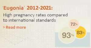
Η Ευγονία διαθέτει  ένα επιτυχημένο και εξειδικευμένο πρόγραμμα δωρεάς ωαρίων, βασισμένο σε αυστηρά πρότυπα ηθικής και δεοντολογίας. Το πρόγραμμα μας εστιάζεται στην ασθενή, αποφεύγοντας το προφίλ προγραμμάτων δωρεάς μαζικής κλίμακας. Αυτό αντανακλάται και στα κορυφαία ποσοστά επιτυχίας της Μονάδας.

Ποιές γυναίκες χρειάζονται δωρεά ωαρίων

Αξιολόγηση και εξετάσεις της δότριας

Νομοθεσία

Διαδικασία δωρεάς

Ποιες γυναίκες χρειάζονται δωρεά ωαρίων

Είναι γνωστό ότι ορισμένες γυναίκες δεν έχουν την δυνατότητα να παραγάγουν δικά τους ωάρια. Οι γυναίκες αυτές δεν έχουν άλλον τρόπο για να τεκνοποιήσουν, παρά μόνον την δωρεά ωαρίου. Αυτό σημαίνει ότι πρέπει να υποβληθούν σε εξωσωματική γονιμοποίηση, κατά την οποία θα χρησιμοποιηθεί το σπέρμα του συζύγου τους, με ωάρια κάποιας άλλης γυναίκας.  Η δωρεά ωαρίων αφορά την διάθεση όλων των ληφθέντων ωαρίων από ανώνυμη δότρια για χρήση από μία ή περισσότερες ανώνυμες λήπτριες.

Στην κατηγορία αυτή βρίσκονται:

  • γυναίκες οι οποίες για κάποιο λόγο έχουν χάσει τις ωοθήκες τους,
  • γυναίκες που έχουν υποστεί ακτινοθεραπεία ή χημειοθεραπεία,
  • νεαρές γυναίκες σε πρόωρη εμμηνόπαυση,
  • γυναίκες των οποίων οι ωοθήκες δεν ανταποκρίνονται ούτε σε ιδιαίτερα υψηλές δόσεις διεγερτικών φαρμάκων.


Αξιολόγηση και εξετάσεις της δωρήτριας

Πραγματοποιούμε ενδελεχή αξιολόγηση όλων των υποψήφιων δωρητριών, και μόνο αυτές που είναι ιατρικά και ψυχολογικά υγιείς γίνονται δεκτές στο πρόγραμμα. Επίσης, καταβάλλουμε κάθε προσπάθεια να αντιστοιχίσουμε τα χαρακτηριστικά της δωρήτριας με τα δικά σας στο μεγαλύτερο δυνατό βαθμό.

Συγκεκριμένα, στην Ευγονία οι δωρήτριες αξιολογούνται με βάση το προσωπικό και το ιατρικό τους ιστορικό από ομάδα ειδικών.

  • Στην πρώτη συνάντηση γίνεται λεπτομερής λήψη ιστορικού από την υπεύθυνη του προγράμματος δωρεών.
  • Κατά τον βασικό έλεγχο από τον υπεύθυνο γιατρό αναπαραγωγής σημαντική προϋπόθεση είναι η εκτίμηση των ωοθηκικών εφεδρειών αλλά και τα επίπεδα των ορμονών.
  • Στη συνέχεια, σε συνεργασία με τον γενετιστή εξετάζεται με λεπτομέρεια το οικογενειακό ιστορικό κληρονομικών διαταραχών και συστήνονται εξειδικευμένες εξετάσεις όπως για παράδειγμα:
    Κυστική ίνωση (έως 99% των μεταλλάξεων του γονιδίου CFTR), ευαίσθητο Χ χρωμόσωμα, α,β και δβ -θαλασσαιμία, νωτιαία μυική ατροφία (εξώνια 7 και 8), μη συνδρομική βαρηκοΐα (μεταλλάξεις 35delG and L90p), καρυότυπος.
  • Επίσης οι δωρήτριες ωαρίων υποβάλλονται σε περαιτέρω έλεγχο,( εξετάσεις για σεξουαλικώς μεταδιδόμενα νοσήματα, προγεννητικό έλεγχο, μοριακοί έλεγχοι για ηπατίτιδες και HIV ) υπερβαίνοντας κατά πολύ τις υποχρεωτικές από το νόμο εξετάσεις.
  • Οι δωρήτριες ωαρίων, καθώς και οι αποδέκτες, έχουν τη δυνατότητα για ραντεβού με εξειδικευμένο ψυχολόγο της Μονάδας, εάν το επιθυμούν.

Τέλος, σημαντική παράμετρος στα προγράμματα δωρεών είναι η πλήρης ενημέρωση των υποψήφιων σχετικά με την διαδικασία της εξωσωματικής γονιμοποίησης, τους πιθανούς κινδύνους και την νομοθεσία.


Νομοθεσία

  • Ανωνυμία: Σύμφωνα με το ισχύον ελληνικό νομικό πλαίσιο είναι υποχρεωτική η διαφύλαξη της ανωνυμίας των δοτών και των ληπτών γαμετών και εμβρύων. Επίσης, τηρείται ένα απόρρητο αρχείο των δωρεών, το οποίο δεν κοινοποιείται ούτε στους δότες, ούτε στους λήπτες, αλλά μπορεί να χρησιμεύσει μελλοντικά, εάν και εφ’όσον κάτι τέτοιο απαιτηθεί διά νόμου. Η ανωνυμία και εν γένει το ιατρικό απόρρητο των δωρεών διαφυλάσσεται απολύτως: εάν έχετε αποδεχθεί την δωρεά ωαρίων ή εμβρύων, δεν θα σας ενημερώσουμε με κανένα τρόπο για την έκβαση της προσπάθειας των ληπτών. Αντιστοίχως, οι λήπτες δεν θα μάθουν κανένα στοιχείο σχετικό με την ταυτότητά σας ή τον ιατρικό σας φάκελο, πλην των πληροφοριών που σχετίζονται άμεσα με την υγεία τους (π.χ. ομάδες αίματος-Rhesus, “στίγμα” Μεσογειακής αναιμίας κ.λπ.).
  • Όρια ηλικίας: Η δότρια ωαρίων πρέπει να είναι 18-35 ετών. Η επιτρεπόμενη ηλικία για υποβολή σε εξωσωματική γονιμοποίηση για όλες τις γυναίκες είναι πριν τη συμπλήρωση του πεντηκοστού έτους.
  • Συγκατάθεση των δοτών και των ληπτών: Η πολιτική μας στον τομέα αυτόν είναι απόλυτη. Θεωρούμε αδιανόητη την δωρεά ωαρίων ή εμβρύων χωρίς την λεπτομερή ενημέρωση και την έγγραφη συγκατάθεση των τεσσάρων εμπλεκομένων: οι δότες καλούνται να υπογράψουν ειδικά έντυπα, με τα οποία εξουσιοδοτούν την θεραπευτική ομάδα να προχωρήσει στην δωρεά οι λήπτες υπογράφουν αντίστοιχα έντυπα, με τα οποία δηλώνουν ότι αποδέχονται την δωρεά. Τα έντυπα αυτά τηρούνται στο απόρρητο αρχείο.
  • Αριθμός μεταφερομένων εμβρύων: Επιτρέπεται η μεταφορά έως 2 εμβρύων, όταν αυτά προέρχονται από δανεικά ωάρια.
  • Διάρκεια κρυοσυντήρησης: Τα κατεψυγμένα έμβρυα μπορούν να διατηρηθούν για χρονικό διάστημα 5 ετών. Η διάρκεια της κρυοσυντήρησης μπορεί να παρατείνεται για πέντε (5) έτη κάθε φορά με έγγραφη αίτηση των δικαιούμενων και ανώτατο όριο παράτασης τα είκοσι (20) έτη (Ν. 4737/2020 (ΦΕΚ Α’ 204/22-10-2020).


Διαδικασία δωρεάς – Λήπτες

1. Αξιολόγηση, ενημέρωση και συμβουλευτική

Στην 1η συνάντηση που θα έχετε μαζί μας, θα συμπληρώσουμε ένα λεπτομερές ιστορικό. Στη συνέχεια θα συζητήσετε με τον Διευθυντή της μονάδας για την προοπτική της δωρεάς, την κατάλληλη θεραπευτική προσέγγιση και το πρωτόκολλο φαρμακευτικής προετοιμασίας αφού γίνει πλήρης κλινική αξιολόγηση.

θα ενημερωθείτε πλήρως για το νομοθετικό πλαίσιο, την προετοιμασία και τις εξετάσεις της δωρήτριας, θα πάρετε πληροφορίες και οδηγίες για την δική σας αγωγή αλλά και συνολικά για την διαδικασία από την υπεύθυνη του προγράμματος δωρεών.

2. Κύκλος προετοιμασίας

Για να μεγιστοποιήσουμε την πιθανότητα επιτυχίας θα υποβληθείτε σε συγκεκριμένες εξετάσεις που θα σας προτείνουμε ανάλογα με το ιστορικό σας.

Αυτές μπορεί να περιλαμβάνουν υστεροσκόπηση, προγεννητικό έλεγχο, εξετάσεις του συζύγου.

Επιπλέον, αν το επιθυμείτε μπορείτε να ζητήσετε ραντεβού με την ψυχολόγο μας. Στόχος μας είναι να βοηθήσουμε το ζευγάρι να εξετάσει και να λάβει υπόψη του όλες τις πιθανές επιπτώσεις που μπορεί να έχει η προτεινόμενη θεραπεία, στους ίδιους, στην οικογένειά και στο παιδί που θα γεννηθεί ως αποτέλεσμα της θεραπείας αυτής.

3. Κύκλος θεραπείας

Είναι σημαντικό για την λήπτρια να έχει προετοιμαστεί με την κατάλληλη θεραπεία ώστε να συγχρονιστεί το ενδομήτριο της με τα γονιμοποιημένα ωάρια. Ο κύκλος θεραπείας για τον συγχρονισμό της προσπάθειας δημιουργεί το κατάλληλο περιβάλλον για την υποδοχή των εμβρύων. Ο κύκλος με ορμονική θεραπεία υποκατάστασης αποτελεί ιδανική λύση . Οι θεραπείες ξεκινούν βάσει περιόδου και περιλαμβάνουν χάπια και ενέσεις.

4. Το εργαστηριακό στάδιο

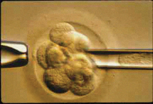
Το εργαστηριακό στάδιο αφορά τη γονιμοποίηση των ωαρίων με το σπέρμα του συντρόφου και την καλλιέργεια των εμβρύων στο εργαστήριο για 2-5 ημέρες.

Η όλη διαδικασία ολοκληρώνεται με τη μεταφορά των εμβρύων στην κοιλότητα της μήτρας της λήπτριας. Υπεράριθμα έμβρυα καταψύχονται και φυλάσσονται στην Τράπεζα Κρυοσυντήρησης της Ευγονίας για μελλοντική χρήση.


Εάν ενδιαφέρεστε να γίνετε δότρια ωαρίων και είστε κάτω των 35 ετών (απαραίτητη προϋπόθεση σύμφωνα με την νομοθεσία),  μπορείτε να επικοινωνήσετε μαζί μας.


Embryo donation is indicated for couples with loss of reproductive capacity (usually both the female and the male partner).

The embryos for donation are provided by a couple who have already undergone in vitro fertilization and have usually completed their family planning. These are surplus cryopreserved embryos, which the donor couple prefers to donate rather than destroy. It is a generous and altruistic act. The decision lies with the donor couple. When they decide to donate, they need to jointly sign a special consent form to declare that they waive all rights and that they are donating the embryos anonymously and without financial compensation to unknown recipients.

According to the Greek legislation, donated embryos must come from a woman under 40 and a man under 50 years old.  The couple wishing to receive embryos shall select, in collaboration with the EYGONIA staff, the embryos that match their characteristics as closely as possible. These embryos are thawed and transferred to the woman following the protocol of a standard frozen embryo transfer with or without hormone replacement therapy (ΗRT). From our very first meetings, we provide the recipient couple with the required consent forms, which they must sign before deciding to receive the embryos.


Under the current Greek legal framework, a program for egg and embryo donation applies under certain conditions:

Safeguarding the recipients' health
Oocyte and embryo donors must undergo a series of clinical and laboratory tests to ensure that no infections will be transmitted to the recipients. All couples that join the IVF program at Eugonia IVF undergo these same checks.

Donation Anonymity
According to the Greek legislation, egg donation refers to the disposal of the retrieved eggs, after the donor's written consent, for use by one or more recipients. In particular, according to Law 4958/2022-FEK142/A/21-7-2022, the identity of the donors, at the choice of these people, can be anonymous, named or disclosed to the resulting offspring after they reach adulthood, upon request. Additionally, recipients can choose whether they want an anonymous or named donor. Donation of reproductive material between relatives is allowed under certain conditions. Finally, the identity of the offspring, as well as its parents (the recipients), is not disclosed to third party donors of gametes of fertilized eggs. According to the current legislation, it is mandatory to keep track of and register all data concerning donors and recipients in the National Donor Registry of the National Authority of Medically Assisted Reproduction, to ensure traceability of the reproductive material.

Age
According to the Greek legislation, the egg donor must be 18 – 35 years old, while the embryo donor must have not exceeded the age of 40.

Compensation
It is prohibited to provide eggs or embryos in exchange for any kind of compensation to the donor.

Ethical side of the donation
Donations, when they are carried out, are driven by necessity, with the "sacred purpose" of the joy of procreation. We believe that before a couple decide to donate or receive donated eggs or embryos, they must give written informed consent. This is why we provide all relevant information from our very first meeting with the prospective donors and recipients. We believe it is our duty to give you all the time you need to assess all relevant information, discuss among yourselves and decide freely and responsibly. Egg and embryo donation is the highest expression of solidarity towards another couple facing the same problem: infertility. Your decision to donate eggs / embryos will by no means affect the course of your treatment.

Consent of donors and recipients
We follow a strict policy regarding consents. We consider it inconceivable to donate eggs or embryos without the written informed consent of all four parties involved: donors are asked to sign a specific consent form authorizing the clinical team to proceed with the donation; recipients sign a corresponding form declaring that they accept the donation. These consent forms are countersigned by a member of the medical team and are strictly confidential.


If you wish to donate your eggs please fill out the form below


In most cases, severe male-factor infertility can be resolved through Intracytoplasmic Sperm Injection (ICSI).

However, in rare cases, sperm may be completely absent from semen (non-obstructive azoospermia). In such instances, the couple can only achieve conception by using donor sperm.

Donor sperm is obtained from specialized sperm donor banks. The minimum standards include the use of cryopreserved semen sample from a donor, who has been thoroughly tested for specific hereditary and acquired diseases, as required by law and international ethical standards. Donor selection is typically based on external characteristics such as hair color, eye color, physique, blood type, and other criteria.

"Eugonia" collaborates with reputable and qualified sperm banks in Greece and abroad, where donors are selected based on strict criteria, both in terms of their fertility as well as their physical and mental health. All samples are accompanied by the test results and reports of the donors.

According to the revised terminology of the International Society for Mild Approaches in Assisted Reproduction (ISMAAR), mild stimulation protocols include a) the natural cycle, b) modified natural cycle, and c) mild stimulation.

Mild ovarian stimulation involves the administration of a low dose of FSH (up to 150 IU/day) for a shorter duration, aiming at the development of fewer follicles and the collection of up to 7 oocytes. Mild stimulation protocols mainly use GnRH antagonists.

However, the use of mild stimulation protocols remains low, mainly because they require a sound knowledge of the GnRH antagonist protocol and optimal laboratory conditions.

At Eugonia we successfully use mild protocols for ovarian stimulation with specific criteria, taking into account the history of each individual couple.

In the field of IVF new knowledge from scientists around the world is added daily, in order to increase success rates. Keeping up to date with new developments is essential.

Our team’s awareness and complete knowledge of the field of fertility is attested by our numerous publications in prestigious scientific journals, oral presentations in scientific conferences and the participation of our scientific team members in European Fertility Societies (ESHRE), besides the international recognition of our researchers.

The exposure to the evaluation and criticism of the scientific community requires us to be constantly updated, as well as to maintain a data-base for the statistics necessary for scientific comparison. These are part of the practice of "evidence based medicine", which is a basic tenant of the Eugonia team.

See our scientific publications

The new generation GnRH antagonists have introduced a new era in ovulation induction. The international medical community tends to recognize GnRH antagonists's protocols as first-line treatment in assisted reproduction, as they offer many advantages such as:The new generation GnRH antagonists have introduced a new era in ovulation induction. The international medical community tends to recognize GnRH antagonists's protocols as first-line treatment in assisted reproduction, as they offer many advantages such as:

  • They are friendly to the patient because they have shorter duration, fewer days of injections, lower doses, lower costs and fewer complications.
  • They are offered for personalized ivf treatment, according to the ovarian response of each woman.
  • They are associated with significantly lower chance of complications (like OHSS) compared to the long protocol, but offer similar pregnancy rates in women with PCOS.Read more in our published study (Lainas et al., 2010 Hum Reprod).
  • They offer significantly higher pregnancy rates in poor responders compared to the short protocol.Read more in our published study (Lainas et al., 2008 Hum Reprod)
  • They are associated with higher pregnancy rates when the antagonist is initiated earlier than the traditional 6th day of stimulation(Read more in our published study Lainas et al., 2005 Hum Reprod).
  • A GnRH agonist can be administered to trigger ovulation, instead of the traditional hCG injection, completely avoiding the incidence of OHSS in high risk women.
  • They are useful on Natural Cycles since premature follicular rupture and oocyte loss are avoided.
  • They can be combined with the single injection protocol of corifollitropin –α (Elonva)

Corifolitropin –a (single injection)


Corifollitropin-a (Elonva) is a new recombinant gonadotrophin (FSH) designed for ovarian stimulation.

The drugs used in ovarian stimulation are of vital importance for the development of multiple follicles and oocytes, aiming at increasing pregnancy rates. The protocols of ovarian stimulation currently used, include daily gonadotrophin injections, combined with GnRH agonist or antagonist, depending on the protocol. Daily injections cause a physical burden in women undergoing IVF treatment.

Elonva is administrated once as a single injection at the beginning of the follicular phase and can sustain multiple follicle development for up to 7 days. From Day 5 of stimulation we start administration of GnRH antagonist to prevent a premature LH surge.

Two large clinical trials have evaluated positively the effectiveness and safety of corifollitropin-a. These studies showed that following a single injection of corifollitropin-a, ongoing pregnancy rates, the number of oocytes retrieved, and side-effects were similar to traditional protocols (i.e. using daily injections). Therefore, a single injection of corifollitropin-a is more patient-friendly and can substitute the unwanted daily injections for 7 days.

Corifollitropin-a is mainly used in a GnRH antagonist protocol. Therefore, knowledge and experience on the use of this protocol is a prerequisite for the administration of corifollitropin-a.

The scientific team of Eugonia has extensive knowledge and experience, as well as a series of published papers on the use of GnRH antagonist protocols. We now participate in an international study to evaluate corifollitropin-a. The first results are encouraging and a significant amount of experience has been accumulated.

During a natural cycle, no drugs are used for the stimulation of the ovaries. The development of the single leading follicle and the maturation of the endometrium are monitored with a series of ultrasounds and hormone tests. Due to a high rate of premature ovulation and loss of the oocyte, the natural cycle has been replaced by the modified natural cycle.

During a modified natural cycle (MNC), antagonist and minimal doses of gonadotrophins are administered in the last days of the follicular phase. In this way premature ovulation is avoided. The advantages of natural cycles are no/minimal drugs, no side effects or complications, short duration, low cost etc.

The disadvantage is that from a single follicle we must retrieve a single oocyte, which must be mature, get fertilized, divide into a good quality, chromosomaly normal embryo with high developmental competence, capable of implanting in the uterus and result in pregnancy.

Indications for natural/modified natural cycle include poor ovarian response, several previous IVF attempts, the desire to avoid drug administration, and some rare contraindications of drug stimulation.

Eugonia - Assisted Reproduction Unit
Konstantinou Ventiri 7(HILTON), 11528 Athens

  • Email: info@eugonia.com.gr
  • Τel.: +30 210 723 6333
  • Fax: +30 210 721 3623

Member of:

Google Reviews

Photo gallery

© Copyright 2024 «Ευγονία» - Μονάδα Εξωσωματικής Γονιμοποίησης στην Αθήνα Ανάπτυξη εφαρμογής NetValue. Με την επιφύλαξη παντός δικαιώματος.
Menu