IVF

Natural Cycle - Modified Natural Cycle (M.N.C)

During a natural cycle IVF there is no ovarian stimulation with drugs; we monitor the development of the single (predominant) follicle and of the endometrium with a series of ultrasound and hormonal tests. The natural cycle itself is a low cost procedure and carries minimal physical suffering. However, because of the occurrence of a high rate of premature and untimely ovulation and egg collection failure, combined with low success rates and a high probability of the cycle being cancelled, this treatment is rarely advised and has been replaced by the modified natural cycle (MNC).

In a modified natural cycle (MNC) low doses of drugs are administered during a natural ovulation cycle. Our goal is to collect one egg, but with a lower chance of canceling the cycle. The addition of drugs in specific days of the cycle, proper monitoring and measurements of the hormone LH that is responsible for premature ovulation, these are all factors that contribute in avoiding cycle cancellation, which has unpleasant consequences for the woman.

The MNC may include the administration of drugs such as the GnRH antagonist to avoid early surge of LH, gonadotropins and hCG to encourage maturation of the egg and support the corpus luteum.

To apply the MNC, deep knowledge and experience of the Antagonist Protocol are vital prerequisites. In Eugonia, we have published a series of studies in valid international scientific journals regarding this protocol and we have extensive experience in implementing it.

This method is the most common version of a natural cycle in IVF. It is advised to women who do not wish to receive high drug doses, while recent studies indicate that the modified natural cycle is associated with higher success rates in women with poor ovarian response. As such, indications for its application include poor ovarian response (not always related to the woman’s age), previous failed attempts, the desire of avoiding medication and contra-indications in pharmaceutical stimulation.

The advantages of natural cycles include the absence of ovarian stimulation, lack of side effects and complications, short duration of the cycle, low cost etc.

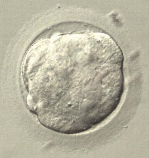
The disadvantages are the availability of only one follicle that we hope will contain one egg, which must be mature in order to be fertilized, it must divide and be able to create a good quality embryo that will be chromosomally normal with good dynamics, capable of being implanted in the uterus and give rise to a pregnancy. Thus, this treatment is considered to be associated with low pregnancy success rates.

Here in Eugonia, the experience and specialization in the MNC have led to considerable pregnancy rates. 

Eugonias Contribution

Our team has published a study supporting that the MNC is related to a four-times higher possibility for live birth compared to the conventional protocol and the use of high dosage of gonadotropins in women with poor ovarian response.

Live birth rates after modified natural cycle compared with high-dose FSH stimulation using GnRH antagonists in poor responders.

Lainas TG, Sfontouris IA, Venetis CA, Lainas GT, Zorzovilis IZ, Tarlatzis BC, Kolibianakis EM.
Human Reproduction 2015;30:2321-30. See the publication

Εξωσωματική γονιμοποίηση τι είναι και πως γίνεται
in vitro fertilization (IVF)

For many couples with fertility problems, in vitro fertilization (IVF) is the best solution in order to achieve a pregnancy. IVF can overcome many causes of infertility, such as mild semen problems, endometriosis, tubal issues or unknown causes. The oocytes are fertilised by the spermatozoa in the laboratory, either by the technique of conventional in vitro fertilization (IVF) or by intra-cytoplasmic sperm injection (ICSI).

IVF History

Robert G. Edwards, became the most famous biologist of his time, when he announced on the 25th of July 1978, Louise Brown’s birth, the first “Test-tube baby” in England, together with obstetrician Patrick Steptoe and the midwife Jane Purdie. Since then, developments in the field have been rapid: in 1992 the first successful fertilization with ICSI was announced (Palermo) as well as its variations, MESA (Silber & Asch, 1992) and TESE (Schoyssman, 1993). In Greece, the first birth via IVF was achieved in 1982 and it was the 13th child was born worldwide.

Stages of IVF treatment- Procedure

Treatment with IVF consists of the following stages:

  • Investigation and infertility diagnosis.
  • Recommended tests before treatment.
  • Ovarian stimulation (12-14 days). We choose the most appropriate protocol based on each woman’s cycle, age and history.
  • Monitoring of the cycle with ultrasound and blood tests every 2-3 days in order to optimize the dosage of the drugs.
  • Induction of the final oocytes’ maturation. It’s the final drug injection that will induce the final maturation of the oocytes and ovulation.
  • Oocyte retrieval (egg collection). It is performed transvaginally, in the presence of an anesthetist. It is short (10-30 minutes) and practically painless due to the light sedation administered.
  • Sperm collection. In cases of azoospermia, testicular biopsy is recommended (TESE)
  • Egg fertilization from the spermatozoa. In this laboratory stage, the oocytes come into contact with the spermatozoa. When semen has normal parameters, conventional IVF is advised. As the the least invasive method for the eggs. For many couples with poor sperm quality, where spermatozoa are unable to fertilize the oocyte unassisted, intracytoplasmic sperm injection (ICSI) is selected. This method allows successful fertilization even in cases of severe oligoasthenoteratospermia. The following day the embryologist checks and records the percentage of normally fertilized eggs.
  • Embryo culture. After fertilization, embryos are cultured in the laboratory for 2 to 6 days, until the blastocyst stage. Proper division of the cells is a basic criterion for their quality.
  • Embryo transfer includes the selection and transfer of the best embryos into the uterus. It is performed two to six days after the oocyte retrieval, using a thin catheter. It is a painless procedure that does not require anesthesia.
  • Pregnancy test (blood test for the determination of β-chorionic gonadotropin hormone, performed 13 days after the embryo transfer)
  • Clinical pregnancy test with a transvaginal ultrasound (4 weeks after embryo transfer).
  • Ongoing pregnancy test on the 12th week of pregnancy, i.e. 10 weeks after embryo transfer.

Optional Procedures (Depending on patient’s history)

  • Embryo or egg freezing Freezing allows conservation of oocytes, sperm and embryos for a prolonged period of time (five years or more). With the application of the new freezing method, vitrification, survival rates and pregnancy success rates are higher than the older method of progressively controlled freezing
  • EmbryoGlue. It is a specialized culture medium containing hyaluronic acid during the embryo transfer process, which aids in the attachment of the embryo to the endometrium.
  • Assisted Hatching is a technique applied in the laboratory and facilitates the hatching of the embryo. It is suggested when the egg’s zona pelucida is thick or hard, in cryopreserved embryos or in women with previous failed attempts

Preimplantation genetic diagnosis (PGD) for the detection of genetic abnormalities and Preimplantation genetic screening (PGS) with the new technique of array CGH or NGS, may be used if considered necessary


Video: What is in vitro fertilization

Read also:

Success rates in ivf

Fertilisation in Natural cycle

Pre-implatation Genetic diagnosis and screening

Embryo Cryopreservation

Oocyte cryopreservation  


Εξωσωματική Γονιμοποίηση

For many couples facing fertility problems, IVF is a safe and effective option in order to achieve a pregnancy.

The process of IVF involves fertilising the woman's oocytes by the husband's sperm in the laboratory (instead of inside the fallopian tubes). Fertilization is achieved either by the technique of conventional in vitro fertilization (IVF) or by intra-cytoplasmic sperm injection (ICSI).

Regardless of your age, If you experiencing fertility difficulties, you should contact us, in order to learn more about the factors that affect infertility and how you can increase your chances of conceiving child.

Individualized treatment, combined with the latest techniques and equipment is the most appropriate approach to achieve a pregnancy.

Together we can make dreams come true. You are in the best hands!

Personalization of the treatment is not always provided, and many IVF centers use the same treatment for all couples. Our philosophy in Eugonia is that every couple undergoing IVF is a distinct case, with a specific medical history and cause of infertility. Therefore, the treatment of infertility must not be identical and rigid for everyone.

It is important to design a different treatment for each couple, with flexibility and adjustment during ovarian stimulation, in order to achieve the best result.
It is thus essential to frequently monitor the woman’s cycle with a series of ultrasounds and blood tests. We appreciate that frequent testing might be inconvenient, but it is very helpful in adjusting the protocol selected and determining the right timing for the last injection prior to the oocyte retrieval. These details can make the difference between a successful and a failed attempt.

Dr Lainas is personally involved in assisting all couples who undergo IVF treatment at Eugonia, by tailoring and monitoring the stimulation protocol for each woman with frequent ultrasound scans and blood tests and giving the proper instructions through the midwives via telephone. He is personally carrying out the majority of oocyte retrievals and embryo transfers, ensuring with his experience the best chances for a positive result and avoiding possible complications, for all couples.

This individualized treatment and attention to detail, combined with the deep Knowledge and specialization of our scientific staff, have led Eugonia amongst the top performing clinics not only in Greece, but also internationally.

In Eugonia, we operate under a rigorous code of ethics, faithfully adhering to the legislation and the strict standards of quality assurance of our services.

In the following pages you can find information on the applicable national legislation regarding IVF, Eugonia’s license by the Greek National Authority of Assisted Reproduction and the quality assurance certifications of our Unit.

In this section you can learn about the core of the legal framework of Medical Assisted Reproduction.

For any further questions, the Legal Advisor of the Unit is at your disposal.


For more information, please visit the Greek National Authority of Assisted Reproduction.


Below you may see the laws, regarding the assisted reproduction (in Greek):

Law 3089| 12/2002

MEDICAL ASSISTANCE IN HUMAN REPRODUCTION


Law 3305| 01/2005

MEDICAL ASSISTED REPRODUCTION APPLIANCE


Law 4272| 07/2014

REGULATION FOR CRYOPRESERVATION- GENETIC MATERIAL DISPOSAL-  ART UNITS/ CRYOPRESERVATION BANKS


Decision 2589| 09/2014

DEFYNING THE NUMBER OF TRANSFERRED EMBRYOS DEPENDIND ON AGE- IUI PRERIQUISITES


A couple's wish to have a baby is an important prerequisite in order to undergo IVF treatment. However, the wish is not enough. It very important that the couples choose the IVF Unit based on objective quality criteria. Which may these criteria be? The quality characteristics, as well as the criteria the couple use to select an IVF Unit (see diagram), have been published in journals of scientific societies (ESHRE, ASRM), and have been the topic of international congress lectures. According to the scientific team of Eugonia, the selection criteria must include:

Scientific excellence

  • High pregnancy rates
  • High quality and range of services
  • Research activity and published studies in international scientific journals of high impact
  • Continuous update on recent developments and use of modern methods based on the principles of evidence-based medicine
  • Reduction of physical burden by using new protocols of short duration

State of the art infrastructure of staff and equipment

  • Scientific staff with deep knowledge and experience,
  • Embryology laboratory with modern equipment, adherence to strict cleaning procedures, detailed protocols, and certification by a government authority.
  • Creation and constant update of a database
  • Quality management certification ISO 9001

Human-centered approach

  • Individualized treatment
  • Direct communication
  • Friendly and warm approach
  • Psychological support
  • Reduction of stress associated with IVF treatment
  • Rapid review of the patient's history and initiation of a new treatment cycle in case of a negative result

Reduction of the cost of IVF treatment

At Eugonia, our attempt to adhere to all the above criteria reflects our high pregnancy rates.


What is In Vitro Fertilization

In vitro fertilization (IVF) is the most commonly used ART method. It was first successfully used in the human in 1978.

Detail of a microscope in the IVF laboratory (EUGONIA archive).

IVF is the fertilization outside the body (fertilization in the glass as the term describes). Therefore, the interaction of eggs and sperm and subsequent fertilization are achieved in the laboratory instead of taking place naturally in the fallopian tubes.

This is a deviation which overcomes certain barriers that prevent fertilization from occurring naturally within the body. However, in most cases, the eggs are the woman's and the sperm are the man's, so the resulting embryos are genetically theirs.


Pregnancy rates in IVF

Eugonia is an ART Unit with stable and top pregnancy rates.

See more:  Pregnancy rates at Eugonia

How is IVF done

The eggs are recovered from the follicles that have developed in the woman's ovaries by the procedure of egg collection. In the laboratory, the eggs come in contact with the sperm inside special dishes with culture medium, in order for fertilization to occur. The fertilized eggs (zygotes) are placed inside an incubator and are cultured under special conditions for 2-6 days, so that the embryos can achieve their first stages of development.

Instead of reaching the uterus naturally, i.e. via the fallopian tube, the embryos are transferred to the uterus by a specialise gynecologist using a thin catheter into which the embryos have been previously loaded by the embryologist. The embryos implant in the endometrium by themselves just like in natural conception. If implantation occurs then a pregnancy will follow.

When to choose IVF

There are absolute and relative indications concerning one or both partners.

Absolute indications are:

  • Lack (following removal) or obstruction of fallopian tubes centrally or peripherally (hydrosalpinges).
  • Lack of spermatozoa (azoospermia) requiring surgical retrieval.
  • Very low number of motile normal spermatozoa (severe oligo-asteno-teratozoospermia).


Relative indications include:


* Rare indications

When in vitro fertilization is undertaken for the first time, especially in cases of unexplained infertility, it can also be used as a diagnostic tool: i.e. a detailed record of gamete behavior, fertilization, embryo development and other parameters is kept. This means that following treatment, the reason of infertility can be established from all these details recorded.

Is IVF something unnatural?

Conventional IVF does not violate the laws of natural conception. Despite certain phobias of the public, IVF takes place in the laboratory without further intervention in the interaction of the gametes, while embryo implantation occurs unaided.

Other versions of conventional IVF, such as ICSI, are indeed intrusive methods, but are used to overcome certain problems, most often related to sperm.

In all cases however, if a pregnancy is achieved, it progresses in exactly the same way as following a natural conception.

Will my child be healthy?

IVF children are as healthy and normal as children conceived naturally, and do not present a significant increase in related and chromosome abnormalities, as shown by several large scale epidemiological studies.

This is confirmed by the fact that more than 1.5 million children have been born to date following IVF, and some of them have already had children of their own.

In Vitro Fertilization risks

In general, in vitro fertilization is a risk free and effective method for a subfertile couple to have their own baby. The worries of the public, especially for those that are not aware, although understood, are almost without any basis.

The risk of pharmaceutical stimulation of ovarian function which is called ovarian hyperstimulation syndrome (OHSS), is thought to appear in its severe form in about 1% of all the women that undertake in vitro fertilization treatments. It is a side-effect that can nowadays be prevented in the majority of cases and can also be effectively managed. The scientific team of Eugonia has already published five original studies for the management of established severe OHSS in internationally renowned scientific journals.

The risk of analgesia/light sedation that is administered during egg collection is comparable to any other risk of a simple surgical procedure.

The risk of developing ovarian, uterine or breast cancer is exactly the same as in the general population, as all large international epidemiological studies show. However, breasts should always be examined, especially in women above 35 years of age.

- A couple is described as “infertile” when systematically attempting to conceive for over a year without success, while being at reproductive age.

- IVF is safe and efficient solution,  that has given the joy of parenthood to thousands of infertile couples.

- IVF is the fertilization outside the body (fertilization in the glass as the term describes). Therefore, the interaction of eggs and sperm and subsequent fertilization are achieved in the laboratory instead of taking place naturally in the fallopian tubes. This is a deviation which overcomes certain barriers that prevent fertilization from occurring naturally within the body. However, in most cases, the eggs are the woman’s and the sperm are the man’s, so the resulting embryos are genetically theirs.

- Fertilization takes place in the embryology laboratory by clinical embryologists and the embryos are transferred back to the uterus by a specialized gynecologist. If pregnancy occurs, its course is identical as after natural conception, and the children born are similarly healthy.

- The use of ICSI (intracytoplasmic sperm injection) gives a solution to nearly all cases of male infertility.

Thanks to ICSI, men with severe problems in sperm numbers and motility can become fathers, while in the past they had no hope of fatherhood. All that is required is a few motile spermatozoa for the fertilization of an equal number of eggs.

- Laser laparoscopic and   hysteroscopic surgery is the method of choice for infertility-related conditions, but is also a modern safe and effective method to treat the majority of benign gynecological conditions.

- The rapid developments that occur daily in the physiology of reproduction, embryology, reproductive endocrinology, laparoscopic and   hysteroscopic surgery offer new means to treat infertility. At the same time, the evolution of technological equipment and the groundbreaking progress in their application have lead to a dramatic increase of pregnancy rates worldwide in state-of-the-art IVF Units.

The IVF treatment cycle includes the following stages:


What must I do first?

You must first book an appointment either yourselves or via one of our collaborating doctors who referred you to our unit. It is also useful that you gather any previous related tests (semen analysis, hysterosalpingography, blood tests, hormone levels, photographs or videos from previous hysteroscopies or laparoscopies), reports from previous IVF attempts, operations, laparoscopies etc.

What tests are necessary?

The preliminary tests before the onset of an IVF program include:

  • Sperm tests (semen analysis, sperm preparation, semen culture)
  • Hormone profiling and transvaginal ultrasound on the 3rd day of the cycle
  • Blood tests for hepatitis, HIV I-II, VDRL (necessary)
  • Pap-test, breast examination in women over 35, heart check up
  • Special examinations, such as laparoscopy or hysteroscopy, which may be recommended by the Medical Director of the unit, based on the assessment of the medical history and other tests (information on laparoscopy-hysteroscopy can be found in a following section).


The tests of prenatal diagnosis are also recommended. These tests usually include haemoglobin electrophoresis (helpful for diagnosis of haemoglobin disorders), blood group, Rhesus factor, rubella, toxoplasm and cytomegalovirus antibodies, complete blood count, complete urine testing, blood urea and sugar, cystic fibrosis etc.

Coloured karyotype (Mr Pagkalos archive)

Although these tests are usually performed in the first weeks of pregnancy, it is preferable to complete them prior to embarking on in vitro fertilization treatment.

In very rare circumstances, a karyotype may be requested, along with a check for thrombophilia and special immunological testing.

Where can I have my tests?

Eugonia covers the entire spectrum of the necessary examinations and collaborates with many national insurance organisations.

It is necessary that the ultrasound, hormone tests and sperm tests are performed in our unit so that the results can be assessed by the scientific team of Eugonia and be compared with similar other cases. This provides an additional level of quality control of the whole process.

However, if this is not possible, these tests can be performed elsewhere and be brought or delivered to our unit. It is preferable that all the tests have been completed before the start of the treatment program.

Monitoring


Ultrasound unit, ADARA type by Siemens.

The monitoring is performed in our unit and involves a series of ultrasounds and hormone tests. You may find a detailed description in the section: Treatment course.


Your first visit at Eugonia

At your first appointment, you will be welcomed by the coordinator of the unit (sister or midwife). The interview will be conducted in three phases:

First, the midwife will take a detailed record of your medical history and information from any previous attempts.

Then, you will meet the Medical Director of the unit, with whom you will discuss extensively the diagnostic and therapeutic strategy recommended. The Doctor will note in your history sheet all the necessary tests that need to be done, the treatment protocol, doses of drugs and details about the treatment program you will follow.

In a third phase, the midwife will explain and give you written instructions for any further tests, if necessary, as well as details about when and how these will be conducted. You will also be given the prescriptions for the necessary drugs, instructions about the treatment protocol, dosage, time and manner to take your drugs, and also medical notes, forms and instructions about your next appointment with us.

You must remember that this first appointment may last a while, depending on the information that need to be recorded and the extent to which you wish to discuss your treatment with the Doctor (usually 1-2 hours).

Finally, you will be given official information brochures and consent forms required by the law, so you have enough time to peruse them. Consent forms must have been signed and filed in your medical folder before the onset of the treatment.

Your visits to the unit during the treatment program

You will be informed about your visits to the unit during the treatment for hormone tests and ultrasounds. Should you have any questions or problems feel free to contact the units midwife, who, in turn, will inform the Doctor.

In each phase of the program, the results of your tests will be assessed by the Medical Director and his colleagues, aiming to decide on the way forward regarding your drug dosage and treatment continuation. Each organism responds differently to the treatment so each case is judged on its own merits. Perhaps it may be possible to re-assess the dosage of the drugs, or modify some timing aspects of the program. Our principle in your monitoring is always the best outcome of your attempt and your final success.

At your first appointment, you will be welcomed by the coordinator of the unit (sister or midwife). The consultation/interview will be conducted in three phases:

First, the midwife will take a detailed record of your medical history and information from any previous attempts.

Then, you will meet the Medical Director of the unit, with whom you will discuss extensively the diagnostic and therapeutic strategy recommended.

The Doctor will note in your history sheet all the necessary tests that need to be done, the
treatment protocol, doses of drugs and details about the treatment program you will follow.

In a third phase, the midwife will explain and give you written instructions for any further tests, if necessary, as well as details about when and how these will be conducted. You will also be given the prescriptions for the necessary drugs, instructions about the treatment protocol, dosage, time and manner to take your drugs, and also medical notes, forms and instructions  about your next appointment with us.

You must remember that this first appointment may last a while, depending on the information that need to be recorded and the extent to which you wish to discuss your treatment  with the Doctor (usually 1 - 2 hours).

Finally, you will be given official information brochures and consent forms required by the law, so you have enough time to peruse them.

Consent forms must have been signed and filed in your medical folder before the onset of the treatment.


Why we use drugs

Drugs used in IVF Treatment
Fertility drugs (Eugonia archive)

It has been shown that pregnancy success rates following IVF increase when more than one good quality embryos are transferred to the uterus. However, embryos formed after IVF do always have the desirable quality. Therefore, in order to have the ability of selection, we need several embryos, which will derive from the fertilisation of several eggs. The eggs, in turn, will be retrieved from several follicles. We therefore use drug protocols of controlled ovarian stimulation to promote the development of multiple follicles.

Why drugs are necessary

The drugs used are analogues of natural hormones in order to create a pharmaceutically controlled reproductive cycle. They aim at the following:

  • The recruitment and maturation of multiple follicles
  • To prevent untimely ovulation and loss of eggs due to early rise of LH. The drugs temporarily disrupt the communication between the pituitary and the ovaries leadin to downregulation of the glands (using GnRH agonists or antagonists)
  • The selection of the ideal time of ovulation (with the application of hCG)
  • To modulate the uterine environment for the support of the embryo (use of progesterone)


Do the IVF drugs reduce my ovarian reserve?

No, as the drugs rescue and promote the maturation of follicles that would otherwise degenerate through atresia. Atresia is the degeneration of follicles initially recruited in a single menstrual cycle.It is worthwhile noting that the ovary contains approximately 300-400.000 follicles, of which only 400 mature during a woman's reproductive life (the rest degenerate via atresia, i.e they become atretic).

Possible side effects of IVF drugs

Mild symptoms from certain drugs and rare allergic reactions (hot flushes, headaches, sweating, blocked nose) are practically insignificant and can be easily overcome.Ovarian hyperstimulation syndrome (OHSS) is a side effect of controlled ovarian stimulation. It occurs in a small percentage of women beginning treatment. In the majority of cases it can be prevented and when it occurs it can be treated.The danger of subsequent ovarian, uterine or breast cancer is similar with the general population as demonstrated by large epidemiological studies.Also see: Ovarian Hyperstimulation syndrome

Which drugs are used?

The main drugs used are:

GnRH analogues

Pharmaceutical analogues of the hormone GnRH intend to inhibit the premature rise of LH that causes ovulation. In this way we are able to prevent the undesirable rupture of follicles before the egg collection. In the past, before the use of GnRH analoguesabout 20-33% of cycles were cancelled due to premature ovulation.GnRH analogues are classified in two categories:

  • GnRH agonists (trade names: Arvecap, Daronda, Suprefact, Gonapeptyl) and
  • GnRH antagonists (trade names: Orgalutran, Cetrotide)

 See: GnRH analogues, GnRH agonists, GnRH antagonists

Gonadotrophins

They are synthetic pituitary gonadotrophins and are applied in order to stimulate the development and maturation of multiple follicles. They are classified in the following categories: 

  • Recombined gonadotrophins (recFSH, recLH)Puregon (recombined FSH)Gonal-F (recombined FSH)Pergoveris (mixture of recombinant FSH, recombinant LH)
  • Urinary gonadotrophins (hMG)Altermon (purified FSH)Metrodin-HP (purified FSH) andΜerional – Bravelle (hMG with FSH-LH mixture)
  • Corifollitropin- α (corifollitropin alpha, with commercial name Elonva)

 See more: Gonadotrophins

Human chorionic gonadotrophin (hCG)

This is the last drug to inject during your treatment. It is applied once at a specific time (36-38 hours before egg collection), when the maturity of the follicles is adequate. Drugs: Ovitrelle (recombined hCG) and Pregnyl, Profasi (urinary hCG)These drugs are available in several forms:

  • ready to use injectable solution
  • powder that is mixed with a special diluent to produce the final solution
  • pre-filled cartridge or pen-like syringe

 Injections are given subcutaneously or intramuscularly, depending on the instructions given. The pen is graduated in international units so you can apply the drug yourselves with precision and safety.See: Human chorionic gonadotrophin (hCG)

Progesterone

It is usually used after the embryo transfer to hormonally support the luteal phase and therefore the environment of the uterus that will receive the embryo. It is available as a vaginal cream (Crinone) or pills (Ultrogestan).Other medicines like estrogens, cortisone, antibiotics and aspirin may be used if deemed necessary. In some cases contraceptive pills are used before the start of the treatment program.See: Progesterone

They are pharmaceutical analogues of GnRH hormone which suppress the premature surge of Luteinising Hormone (LH) that triggers ovulation. In this way, premature ovulation prior to oocyte retrieval is prevented. Prior to the use of GnRH analogues, there was a 20-30% cancellation rate of treatment cycles due to premature ovulation.

GnRH hormone is produced in the hypothalamus area and results in the production, storage and release of gonadotropin hormones FSH and LH from the pituitary gland. GnRH is released periodically (in waves) every one to three hours, depending on the phase of the cycle and is then followed by the release of the gonadotropins from the pituitary gland.

Why do we use GnRH analogues

It is well established that the rise and surge in LH results in the final oocyte maturation and ovulation. In IVF treatment we prefer to schedule ovulation at a specific time. A premature surge in LH would result in follicle rupture prior to egg collection and loss of the ovulated oocytes. The control of LH surge by its suppression is of great importance and has significantly reduced cycle cancellation. This control is achieved through the use of GnRH analogues.

The action of GnRH agonist analogues is similar to that of the natural hormone.

The GnRH agonist analogues, at the start of their administration and also in the case of their intermittent use, result in the stimulation and rise in the release of FSH and LH gonadotropins, through a flare-up mechanism. This happens because they stimulate the synthesis of GnRH receptors in the gonadotropic cells of the hypophysis (pituitary gland).

If the administration of GnRH agonists is prolonged, the ovarian function is suppressed due to the reduction of gonadotropin release. This reduction occurs due to the extended occupation of the GnRH receptors by the GnRH analogues and also due to the reduction of the number of GnRH receptors in the gonadotropic cells of the hypophysis. This phenomenon is also known as down-regulation or pituitary desensitization and has been considered a "paradox" in its first publication in 1975. Nowadays, we are taking advantage of this "paradox" to achieve suppression of the pituitary function by GnRH agonist analogues administration.

When are they used

GnRH agonists are administered towards the end of the last menstrual cycle (in the middle of the luteal phase, meaning around 7 days before the menstrual cycle is due) or in the beginning of the menstrual cycle and they progressively result in the suppression of the pituitary function. Since the pituitary gland is no longer active, the normal course of the menstrual cycle is suspended, the follicles do not mature and they do not rapture, and this allows us to administer gonadotropins in such doses to stimulate multiple follicle development in the time that we chose. Nonetheless, the continuous administration of GnRH analogues is obligatory, during the entire stimulation phase.

Commercial names

GnRH agonists that are available in Greece have the following commercial names: Arvekap (Triptorelin), Daronda (Leuprolide), Suprefact (Buselrelin) and Gonapeptyl. These come either in a pre-mixed dilution ready for subcutaneous injection, or in the form of a powder that needs to be mixed with a special diluent in order to become an injectable solution, or in a nasal spray. Their active ingredients are all similar. GnRH agonists administration may result in a little uterine bleeding (pseudo-period). Possible side-effects of treatment with GnRH agonists are a stuffy nose, hot flashes, headaches, night sweats, etc.

The primary mechanism of action of GnRH antagonist analogues of is to compete with the natural GnRH hormone. GnRH antagonists bind to the GnRH receptor which causes a suppression from the very beginning of their administration, due to the occupation (and not a reduction in the numbers) of GnRH receptors. They thus result in the reduction of FSH and LH gonadotropin production. The suppression of the pituitary gland function is achieved in minimum time (almost straight after) following their administration.

The pharmaceutical production of GnRH antagonist analogues occurred after that of GnRH agonist analogues. After setting up the daily GnRH antagonist dosage, five multicenter phase III studies followed (2000-2001) in Europe, Middle East and USA, in order to extract conclusions for their effectiveness in clinical practice, the comparison of their results with GnRH agonists, etc. Since then, numerous original studies and meta-analyses have been published with regard to the experience of administering GnRH antagonists on a preselected day (typically, the 6th day of stimulation) and the so called flexible administration protocol.

When are they used

Antagonists are administered during the treatment with gonadotropins, in the current menstrual cycle. The main innovation of antagonists is that their administration, even for a few days prior to follicular rupture, causes an immediate suppression of pituitary function that results in the prevention of the LH surge.

Commercial names

GnRH antagonists are available with the following commercial names: Orgalutran (Ganirelix) and Cetrotide (Cetrorelix), in a pre-mixed dilution ready to be used by subcutaneous injection.

It is well know that pituitary hormones FSH and LH are called gonadotropins because they act on the gonads (testicles-ovaries) and promote their function. Nowadays, synthetic and pharmaceutically produced pituitary gonadotropins are available which are administered for the development and maturation of multiple follicles. The drugs that are available are the recombinant and the urinary gonadotropins.

These hormones are administered by injection and they are commercially available:

  • in a pre-mixed ready to used solution,
  • in a powder that needs to be mixed with a special diluent in order to produce the injectable solutio
  • pre-filled cartridge or pen-like syringe.


The pen-like syringe is marked in units so as the woman can administer subcutaneously the solution by herself with precision and safety.

Recombinant gonadotropins

They are new reproductive era drugs. Molecular biology has allowed pharmaceutical companies to apply the developments in recombinant DNA technology in drug production. Two forms of the human FSH gene have been isolated and they have been inserted and integrated in genetically modified cells. These cells can be cultured industrially and can therefore produce and release large quantities of natural human FSH, which can then easily be isolated in an almost pure chemical form.

Nowadays, these gonadotropins are available commercially under the generic name "recombinant FSH". Their commercial names are Puregon and Gonal-F. The chemical purity of these products allows their subcutaneous administration but the can also be administered intramuscularly. The way they are administered does not affect their biological action. The other gonadotropin called LH is also available as a product of the same technology with the commercial name Luveris.

Urinary gonadotropins

They are "older" technology drugs. For many decades, gonadotropins that were used in assisted reproduction came from the urine of postmenopausal women. There was a complicated and expensive system in place for the collection of the urine (usually from women monasteries) and the isolation of hormones, a true struggle of the pharmaceutical industry of which little was known to the general public. With the sudden increase in gonadotropin demand due to the wider application of assisted reproduction, this system quickly reached its definite capacity, in the beginning of the 90's.

The drugs of that era contained a mixture of the two gonadotropins (FSH and LH) in various doses, with the generic name hMG (human menopausal gonadotropin) and were commercially available with the names Pergonal, Humegon, Pergogreen, Humegon FD. Their administration had to be done intramuscularly, due to their rather small grade of chemical purity. During the middle of the 90's, technology evolved and new drugs that contained purified FSH (Metrodin-HP) which were possible to be administered subcutaneously become commercially available. Nowadays, there are still urinary gonadotropins available for intramuscular or subcutaneous injection with the commercial names Altermon, Metrodin-HP and Menogon. Altermon is highly purified product and Menopur, Merional and Bravelle contain a mixture of FSH/LH in a 1:1 ratio.

Corifollitropin-α is a new recombinant gonadotropin (FSH) that has been designed for ovarian stimulation. It is administered in a single injection in the beginning of the ovarian stimulation and can initiate and sustain multiple follicular development for up to 7 days. Also see: The new drug Corifollitropin-a

This is the last injectable drug of the treatment. It is administered in a single dose at a specific time (around 36-38 hours prior to egg collection) when the follicular development is deemed satisfactory to proceed with the egg collection.

hCG is a hormone that is normally secreted during the placenta development. Its measurement is used for the detection of pregnancy. However, an area of the hCG molecule is very similar to the LH molecule and it is therefore possible for the hCG to bind to the LH receptors and mimic its action. This means that the administration of hCG in a single powerful dose can "trick" the ovary and induces follicle rapture around within the next 36 hours (please note that the normal hormonal signal of ovulation in the human body is LH, which is released in a large surge when oestradiol levels go over a certain point).

hCG is available in a pharmaceutical form for intramuscular injection with the commercial names Pregnyl or Profasi (urinary hCG). During the last few years there is also a recombinant form of hCG (r-hCG), with the commercial name Ovitrelle. Ovitrelle is available in a pharmaceutical form for subcutaneous injection due to its high purity.

It is usually used after embryo transfer to hormonally support the luteal phase of the menstrual cycle. According to the literature, the luteal phase of the menstrual cycle needs progesterone support, especially in treatment cycles that a GnRH analogue has been used.

This hormone is available in the form of a vaginal gel (Crinone), or in pills (Utrogestan). These pills may be administered orally, vaginally (as suppositories), or both orally and vaginally. The local absorption of progesterone from the vagina is very good and has proven to have excellent results.

During the course of IVF treatment, the following drugs may also be used.

Antibiotics

The male partner receives prophylactic administration of antibiotics when the female partner starts her ovulation stimulation and the female partner receives prophylactic administration of antibiotics following egg collection, according to the unit's instructions. Vibramycin is usually the antibiotic used.

Corticosteroids

In special cases, drugs that contain cortisone may be used (Medrol pills or Dexamethasone). In a recently published study of our unit, that had a very good response in the international scientific press, it was shown that the use of corticosteroids is related with the prevention of ovarian hyperstimulation syndrome (Administration of Methylprednizolone to prevent severe ovarian hyperstimulation syndrome in patients undergoing in vitro fertilization.) T. Lainas et al., Fertil. Steril. 2002;78(3):529-533). See the publication

In every case, special care should be taken and the doctor's instructions should be followed to the letter when starting and especially when stopping cortisone administration, which should be done by gradually reducing the dose, according to a specific time-plan.

Oestrogens

In certain cases, for instance when the protocol of preparing the endometrium for embryo transfer without having an egg collection is used, e.g. in the case of a frozen embryo transfer, oestradiol is administered orally (Estopause, Cyclacur; only the white pills in the package contain 17- β oestradiol) or with transdermal patches (Dermestril or Estraderm).

Clomiphene citrate

It is an "older" drug with proven results, that has anti-oestrogen action. This drug is used in certain ovarian stimulation protocols, either on its own, or in combination with gonadotropins. Its anti-oestrogen action "tricks" the pituitary gland into thinking that there is no oestrogen available, which results in an increase in the release of endogenous FSH and development of the follicles.

Clomiphene is available in pills, with the commercial names of Clomiphene citrate, Serpafar and Clomid. If the stimulation is relatively mild, clomiphene is mainly used in ovulation induction or in intra-uterine insemination cycles and also in some other rare assisted reproduction treatments.

Schematic representation of fertilization.

Conventional IVF

This represents the main laboratory stage of your attempt. A few hours following the egg collection the embryologist places a specific number of motile spermatozoa in the culture dishes containing the eggs.

In conventional IVF there is no further intervention.

The spermatozoa approach the egg on their own and one of them penetrates it and fertilizes it.

VIDEO: In vitro fertilization (IVF)

In more detail, the spermatozoa digest the layers of granulosa cells, meet the oocyte, attach on the zona pelucida, and only one sperm enters the oocytes.

The oocyte reacts to the penetration of the spermatozoon. First, the zona pelucida and cell membrane change their consistency to prevent the entrance of more sperm. Second, the oocyte resumes the second meitic division and extrudes the second polar body. Simultaneously, the male and female pronuclei form and become visible, marking the fertilization of the oocyte.

Morula.
Morula.

The morula is observed on day 4 and is formed after the compaction and fusion of blastomeres of the dividing embryos. As the morula continues to develop, fluid starts to accumulate forming a cavity, the blastocoel, giving rise to the blastocyst.

Blastocyst

Expanded blastocyst.

The blastocyst forms after 5 days of embryo culture. The mature blastocyst is characterized by expansion of the blastocoel due to accumulation of fluid, the thinning of the zona pelucida,and the formation of two distinct cell layers, the inner cell mass, that will form the embryo, and the trophectoderm, that will form the placenta.

The blastocyst for the evaluation of embryo viability

Development to the blastocyst stage is a sign of normal development and high developmental competence. It has been shown that embryos reaching the hatching blastocyst stage have double chances for implantation. At Eugonia, blastocyst formation rate is 54%. This high percentage reflects optimal laboratory conditions and experience of the scientific staff.

Activation of the embryonic genome occurs between 4 and 8 cells. Therefore, the contribution of the paternal DNA and the assessment of an embryo as a whole can be better performed 5-6 days after oocyte retrieval. This is particularly important for cases with poor sperm quality (oligoasthenozoospermia) or testicular biopsy.

Continuous development of culture media and laboratory conditions allow the development of embryos in vitro for up to 6 days without compromising their viability. However, there is a theoretical chance that the uterus may provide a more favorable environment for embryo development. Practically, however, in an IVF laboratory with high experience and quality control, embryos of higher developmental competence will grow to the blastocyst stage, providing the Embryologist a strong criterion for embryo selection. For this reason, pregnancy rates after transfer of blastocysts is higher and reaches 72% for women of all ages at Eugonia.

These protocols refer to the pharmaceutical regulation in order to achieve controlled ovarian stimulation (COS). The reason for their use is multiple follicle development and avoidance of early follicle rupture due to the surge in LH. Early follicle rupture is equivalent to the loss of oocytes prior to their scheduled pick-up (oocyte retrieval) that is performed by the gynaecologist under ultrasound guidance.

The decision of which stimulation protocol to use depends on the individual nature of each patient's cycle, the ovarian response in previous cycles, the age and other factors assessed by the doctor.

Nonetheless, the application of these protocols is flexible as every patient may respond to the drugs differently. An important part of our task is to define the best therapeutic protocol and decide on the ideal dosage for each separate case.

The most common protocols are:


Long protocol

The long protocol has been in use for quite some time. The first publication was in Lancet scientific journal by Porter and colleagues in 1984 and it is also known as the GnRH agonist down regulation protocol. The basis of this protocol is the down regulation of the pituitary and therefore the prevention of a premature LH surge. It is well known, that a premature LH surge would result in follicle rupture prior to the egg collection and thus in loss of the oocytes.

Pituitary down regulation is achieved with the continuous administration of GnRH agonist analogues. Having established a continuous suppression of the pituitary, the stimulation of the ovaries whose main goal is the recruitment and development of multiple follicles then follows. This means that the communication between the pituitary and the ovaries is cut off and that we are the ones to take over.

The long protocol involves two phases:

  • 1st phase: Downregulation using GnRH antagonists (Arvekap, Daronda, Suprefact, Gonapeptyl). Duration approx. 10-14 days.
  • 2nd phase: Ovarian stimulation using gonadotrophins (Puregon, Gonal-F, Altermon, Pergoveris, Merional, Menopur, Bravelle) during continuous downregulation of the pituitary. Duration approx. 10-14 days.


The total duration of the long protocol is about 1 month.

Onset of downregulation in the long protocol
Downregulation of pituitary and ovarian function is checked at the end of the 1st phase. You may be informed of the exact date of your downregulation check you may contact the midwives of our unit either on the day of analogue administration or on the first day of your period, so they can schedule your next appointment. The downregulation check usually involves a transvaginal ultrasound and measurement of certain hormones. If downregulation is sufficient you will be given instructions to start the gonadotrophins.

Onset of stimulation in the long protocol
We will contact you in the late afternoon of the downregulation day to confirm the completion of your downregulation and the start of your stimulation. You will be given instructions on the dose, start date and times to take your gonadotrophins. Meanwhile, you will continue to take the GnRH analogue until the end of the treatment at the dose suggested by us.

Onset of downregulation in the long protocol

The administration of the GnRH agonist (Arvekap, Daronda, Suprefact, Gonapeptyl) can start:

  1. on the 21st day of the cycle, in a normal 28 day menstrual cycle,
  2. on the 2nd day of the cycle,,
  3. three days prior the end of the treatment with contraception pills.


Right ovary during downregulation. The ovary appears without any cysts during a transvaginal ultrasound (EUGONIA archive).
Downregulation check. The endometrium appears thin during a transvaginal ultrasound(EUGONIA archive).

Downregulation check during the long protocol

Downregulation of pituitary and ovarian function is checked at the end of the 1st phase. You may be informed of the exact date of your downregulation check you may contact the midwives of our unit either on the day of analogue administration or on the first day of your period, so they can schedule your next appointment.

The downregulation check usually involves a transvaginal ultrasound and measurement of certain hormones. If downregulation is sufficient you will be given instructions to start the gonadotrophins.

Onset of stimulation in the long protocol

Long protocol starting on the 2nd day

In this protocol, the administration of the GnRH agonist starts on the 2nd day of the cycle. In this case, there is an initial is stimulation, followed by suppression of the pituitary function, due the GnRH agonist mechanism of action. As a result of this initial stimulation of the ovary, there is a 15-20% chance of a functional ovarian cyst appearing.

Functional cycst of the right ovary (transvaginal ultrasound) (EUGONIA archive).
Transvaginal aspiration of a functional ovarian cyst by ultrasound guidance (EUGONIA archive).

During the check of downregulation, increased levels of oestradiol are also observed. If an ovarian cyst appears, it is aspirated transvaginally by ultrasound guidance and it usually subsides after few days, as the GnRH agonist administration continues. Downregulation check is then repeated after one week and the treatment continues after confirming downregulation (by ultrasound and blood eostradiol level measurement).

Long protocol starting on the 21st day

In this protocol, the administration of the GnRH agonist starts in the middle of the luteal phase of the cycle. If the cycle is regular (28 days), the start date coincides with the 21st day of the cycle. In the case of an irregular cycle, the ovulation needs to be determined and the start of the GnRH agonist administration is scheduled seven days after that, once a progesterone measurement has been performed in order to confirm that we are indeed in the middle of the luteal phase of the cycle.

Long protocol under contraceptive

In the long protocol under contraceptive, the GnRH agonist administration starts three days prior to stopping the contraceptives (3 pills before the end). This protocol is usually suggested in cases of irregular menstrual cycles, polycystic ovarian syndrome, or in cases of ovarian cysts etc.


The advantages of the antagonist protocol

It has been quite some time that the focus of the pharmaceutical companies that specialize in the production of fertility treatment drugs has shifted towards the production of drugs that could provide immediate suppression of LH and quick reversibility of that effect. GnRH antagonists met these specifications, although their safety and efficacy had to be proven in everyday clinical practice.

Nowadays, following numerous published studies and metanalyses that followed the first five multicentre phase III studies (2000-2001), we can say that antagonist protocols are patient friendly because:

  • They have fewer injections
  • smaller treatment duration (at least 12-15 days less when compared to the long protocol)
  • smaller gonadotropin dosage in total
  • less side effects (hot flashes, night sweats, nervousness, insomnia etc.)
  • smaller chance of OHSS (statistically significant)
  • Are suitable for modified natural cycles (M.N.C.)
  • Are suitable for the application of mild stimulation protocols


Antagonist protocols demand a long learning curve and keeping up to date with all the latest studies, which could explain their initial smaller acceptance by the international scientific community.

The scientific team of Eugonia has proven its everyday knowledge and experience in the antagonist protocols, with original studies published in internationally accepted scientific journals. The effort of the scientific team of Eugonia is internationally recognised as a significant contribution to the assisted reproduction field.

Application of the antagonist protocol

In this protocol, gonadotrophic stimulation begins on the 2nd or 3rd day of the cycle, while the downregulation using the antagonist GnRH analogue follows. The duration of the antagonist protocol is approximately 12-15 days.

In the gonadotropin and antagonist protocol, the ovary receives two consecutive stimulation signals: initially from the increased endogenous FSH in the beginning of the cycle, and then from the exogenous injected gonadotropins. This means that follicle recruitment does not cease from the normally increased endogenous gonadotropins at the beginning of the cycle and that the gonadotropins administered from the beginning of the cycle reinforce follicle recruitment.

The antagonist administration can start either blindly on the 6th or the 1st day of stimulation with gonadotrophins (fixed antagonist protocol), or based on ultrasound and hormone criteria (flexible antagonist protocol).

The contribution of Eugonia to the antagonist protocol

The scientific team of Eugonia has published numerous studies on women undergoing assisted reproduction using the antagonist protocol in internationally renowned scientific journals. These are:

Women with polycystic ovarian syndrome

Comparison of long versus flexible antagonist protocol

Our study is the largest one internationally. It is a randomized prospective study that compares long with flexible GnRH antagonist protocol.

The results show that pregnancy rates are similar in both protocols and there is also a significant reduction (by 20%) of severe ovarian hyperstimulation syndrome when the antagonist protocol is used.

The antagonist protocol is suggested as a protocol of choice for women with polycystic ovaries. The conclusions of this study are adopted by independent editors of international organizations (Faculty of 1000 Medicine) and are honorary selected in "Editor's choice" by Professor Andre Van Steirteghem, who is the editor of scientific journal Human Reproduction.

The flattering comment that the suggested protocol can change the routine clinical practice refers to the finding that the flexible antagonist protocol is the safest protocol in women with polycystic ovaries without reducing their pregnancy rates.

See 1000 Medicine
See Editors choice
See our publication

Comparison of long versus antagonist protocol (D1)

In this study, the long protocol is compared with an alternative antagonist protocol. In this antagonist protocol, the first day of antagonist administration is on the 1st day of stimulation instead of the standard day 6 of stimulation./p>

In this original randomized prospective study the hormone levels are examined, along with the follicle development and the differences between the two protocols are highlighted. The study shows the differences in the hormonal environment during the stimulation and that the fastest follicle development happens with the antagonist protocol. This study has a great theoretical interest in the effort of optimizing the antagonist protocols/p>

The results show similar pregnancy rates for both protocols and reduced length of stimulation duration and reduced appearance of OHSS with the antagonist protocol.

See our publication

Poor responders

In our study on poor responders we achieved significantly higher ongoing pregnancy rates using the flexible antagonist protocol, as compared to the short agonist protocol. This is by far the largest randomized controlled trial in the literature on poor responder patients undergoing IVF treatment, and has been included in several meta-analyses.

See our publication

Antagonist administration according to individually selected criteria increases pregnancy rates

When antagonist administration is applied according to individually selected criteria, this correlates with statistically increased pregnancy rates. A relevant study of the Eugonia scientific team was recently published in scientific journal of the European Society of Human Reproduction and Embryology (ESHRE) titled «In a flexible antagonist protocol earlier, criteria-based initiation of GnRH antagonist is associated with increased pregnancy rates in ΙVF» (Lainas et al., Hum Reprod 2005;20(9):2426-2433).

See our publication

The administration of antagonist in the luteal phase can manage already established severe OHSS

See our publications
Live births after management of severe OHSS by GnRH antagonist administration in the luteal phase.

Management of severe OHSS using GnRH antagonist and blastocyst cryopreservation in PCOS patients treated with long protocol.

Management of severe early ovarian hyperstimulation syndrome by re-initiation of GnRH antagonist.

Corifollitropin is a new drug developed for controlled ovarian stimulation, mainly used in a GnRH antagonist protocol. One single subcutaneous injection on the 2nd day of your menstrual cycle can replace a whole week (7 days) of daily FSH injections. On the 5th day of your menstrual cycle your doctor will prescribe you an antagonist. You may need to continue treatment with extra FSH injections until enough follicles of the adequate size are present.For further details click here

It is recommended as an alternative to patients with particularly poor response to ovarian stimulation, such as patients with several previous failed attempts and embryos of bad quality. The development of the follicle and the endometrium is monitored with a series of ultrasounds and hormone measurements. The disadvantage of the natural cycle is that we place our hopes in one follicle from which we have to retrieve one egg that needs to be mature and fertilize normally, divide on time and create an embryo of a satisfactory quality to proceed with an embryo transfer. Of course, in the natural cycle, there is also a chance of a premature LH surge and follicle rupture that would lead to failed oocyte retrieval.

The modified natural cycle is recommended in cases of extremely poor response to ovarian stimulation and also as an alternative in women with multiple failed attempts and in women that do not wish to take any drugs for stimulation. It may also be an alternative solution before oocyte donation is suggested in selected cases.

The scientific team of Eugonia has many years of clinical experience and the know-how in modified natural cycles and has to present an encouraging number of cases with successful pregnancies.

In the short protocol the GnRH agonist is administered almost concurrently with the gonadotropins and it is also known as the Flare-up GnRH agonist protocol. The short protocol is usually selected for women with poor response to ovarian stimulation. In the short protocol:

  • The administration of GnRH agonist usually starts on the 1st-2nd day of the cycle and the gonadotrophins on the 2nd-3rd day.
  • It is not divided into a downregulation and stimulation phase,
  • its duration is about 10-14 days in total.


In the short protocol, with the simultaneous administration of GnRH agonists and gonadotropins we are taking advantage of three stimulation signals of the gonadotropins on the ovaries in order to recruit multiple follicles and to collect more oocytes. These three "triggers" are applied by the increased endogenous FSH that normally increases in the beginning of the cycle, by the increase of endogenous FSH due to the initial stimulating effect of the GnRH agonist in the pituitary and by the exogenously administered gonadotropins via injections. Towards the last days of the short protocol, the suppressive effect of the GnRH agonist in the pituitary also takes place. Variations of the short protocol include the ultra-short and the Microflare (Microdose flare) protocols.

These protocols are rarely used and only in special cases. In these protocols, which are usually used in women with particularly poor response to ovarian stimulation, the GnRH agonists are not used and there is a risk of a premature LH surge. In such instances, a GnRH antagonist may also be used. The start date and the dosage of the drugs administered are set by the doctors of our Unit.

Ultra short protocol

The difference of this protocol when compared to the usual short protocol is in the very short duration of the GnRH analogue administration (about three days). In this way, only the stimulatory action of the analogue on the pituitary (which results in an increase in endogenous FSH) is used and not its suppression effect.

Clomiphen-gonadotropin protocol

In the clomiphene-gonadotropin protocols, clomiphene citrate is usually administered from the 2nd to the 6th day of the cycle, and then the gonadotropin administration follows

ΜΑP

In the MAP protocol a contraceptive pill is used in the previous menstrual cycle. The GnRH agonist administration starts on the 2nd day of the cycle in microdoses. The administration of gonadotropins starts on the 3rd day of the cycle.

See more: Mild stimulation protocols

The transfer of frozen thawed embryos may be scheduled in a pharmaceutically controlled cycle or in a natural cycle by monitoring ovulation.

Pharmaceutically controlled cycle

In this protocol, only the endometrium is prepared with drugs for the embryo transfer. Initially, the pituitary function is suppressed in order to prevent a premature LH surge and therefore ovulation, which would result in the release of progesterone and its effect on the endometrium at the wrong time. A cycle with hormonal replacement therapy would be the best solution.

Suppression starts on the 21st or the 2nd day of the cycle, as with the long protocol, the same steps of controlling the suppression are followed. Stimulation of ovarian function is not necessary. During the 2nd phase of the treatment, the endometrium is prepared with pills or patches of 17-β oestradiol (white pills of Estopause or Cyclacur and Dermestril Estraderm patches. Monitoring is performed by vaginal ultrasound, during which the thickness and the structure of the endometrium are checked. At the end, progesterone pills are also administered and the day of embryo transfer is scheduled.

Natural cycle

The transfer of frozen thawed embryos can be performed in a natural cycle. In this case, the development of the follicle is monitored along with the endometrium with a series of transvaginal ultrasounds and hormone measurements. Ovulation is predicted or induced and the day of embryo transfer is scheduled.

In the IVF treatment programs, the term "poor responder" is commonly mentioned. This means that the ovaries of these women show significantly reduced response to the ovarian stimulation drugs that are administered for the recruitment and development of multiple follicles. When compared with the population of women that show normal ovarian response, poor responders produce fewer eggs and have lower pregnancy rates in IVF treatment cycles.

Unfortunately, there is no internationally accepted definition of poor response. The majority of doctors agree that this cohort, includes women that during an IVF cycle recruited and developed less than 3 or 5 follicles and produced less than 3-5 mature eggs even when high gonadotrophin doses and various ovarian stimulation protocols were used.

Various researchers have described numerous criteria, such as: low E2 levels during stimulation, increased FSH levels, low levels of AMH on the 3rd day of the menstrual cycle, at least one cancelled cycle, no response to increased gonadotropin doses, long stimulation duration etc.

High gonadotropin doses (>300IU) exclude the chance of a reduced dosage that may be linked to the small number of follicles that will be recruited and develop.

Another result of the small number of follicles that develop in the ovaries is the low E2 levels during stimulation.

Poor response is due to reduced ovarian reserve. The ovaries are unable to produce more eggs than the ones they already have, even if they are bombarded with high gonadotropin doses.

It is self explanatory that from a small number of follicles, we will retrieve a small number of eggs. If for example the number of eggs collected is 5, only a percentage of these will be mature (i.e. able to fertilise). Only a percentage of these mature eggs will fertilise, divide and have a good implantation potential.

Such loses significantly lower the number of embryos available for transfer. For this reason, poor responders show lower pregnancy rates when compared to women with a normal response.

The factors responsible for poor response may be genetic and environmental, as well as ovarian surgery, endometriosis, immunological, auto-antibodies, reduced number of FSH receptors on granulosa cells, unknown, etc. Ofcourse, the main factor responsible is a reduced ovarian reserve.

Poor responders are not necessarily subfertile when there is no other cause of subfertility.

Prediction of poor response is possible with: a) a basal antral follicle count (e.g. <5 basal antral follicles) by transvaginal ultrasound, b) increased FSH levels (e.g. >12) on the 3rd day of the cycle, c) low AMH levels (e.g. <0.3-0.6 ng/ml). In the past, inhibin-B and GnSAF were used. In IVF treatment, it is especially useful to record the ovarian response to high gonadotropin doses (>300IU) in any previous cycles.

Management of poor responders

Unfortunately, international scientific literature (Tarlatzis, Ubaldi) shows that there is no ideal stimulation protocol.

There have been described and compared: high gonadotropin doses (>300IU), the use of recombinant gonadotropins versus urinary gonadotrophins, administration of gonadotropins in the luteal phase, short and ultra short GnRH agonist protocols with a reduction in the GnRH agonist dose (Arvekap, Daronda, Suprefact), long protocol with agonist administration in the luteal phase, GnRH antagonist protocol, growth hormone (GH) administration, gluco-corticoids administration, use of contraceptives, aromatase inhibitors, DHEA (dehydroepiandrosterone), use of only intracytoplasmic sperm injection (ICSI) for fertilization, assisted hatching, natural cycles, modified natural cycles (M.N.C.) and finally oocyte or embryo donation

Our contribution for poor responders

We have published the largest study in the international literature for poor responders that underwent IVF treatment. Our study was published in the internationally acclaimed scientific journal of Human Reproduction, which is the official journal of the European Society for Human Reproduction and Embryology (ESHRE). It is a prospective randomized study that shows that the flexible GnRH antagonist protocol correlates with statistically higher ongoing pregnancy rates when compared to the short GnRH agonist protocol. In conclusion, the antagonist protocol may be considered as the protocol of choice for women with poor ovarian response that undergo IVF treatment.

See our publication

This category includes women that show an excessive response to ovarian stimulation drugs which results to an excessive recruitment and multiple follicle development.

The factors that favour excessive ovarian response are polycystic ovaries, young age, reduced body weight (low BMI; body mass index), high gonadotropin doses or progressively increasing dose, aggressive stimulation protocols, excessive ovarian response in a previous IVF treatment cycle, etc.

Hyperesponders have increased chances of developing OHSS (ovarian hyperstimulation syndrome) and possibly the severe form of the syndrome, which is one of the most serious complications of IVF treatment.

The scientific team of Eugonia has shown international innovation and experience in the prevention and management of OHSS with a significant number of studies published in internationally acclaimed scientific journals and lectures of the Head of the Eugonia Unit, Dr T. Lainas.

The publications reflect the knowledge and experience applied in everyday clinical practice.

Prevention of OHSS

The publications related to OHSS prevention are either on corticoids administration (also an older opinion of our Unit), or on the use of antagonist protocols which are thought to be safer for hyperesponders.

Also see: Antagonist protocol.

See our publications:

Live births after management of severe OHSS by GnRH antagonist administration in the luteal phase.

Management of severe OHSS using GnRH antagonist and blastocyst cryopreservation in PCOS patients treated with long protocol.

Management of severe early ovarian hyperstimulation syndrome by re-initiation of GnRH antagonist.

Management of OHSS

The proposal of the scientific team of Eugonia for antagonist administration in the luteal phase for the management of already established severe OHSS is a world's first.

See: our news

The term ovarian reserve refers to the reserves of the ovaries in follicle numbers, which is expressed with the ability of the ovaries to recruit follicles when they are stimulated with drugs. As the age advances, the ovarian response to exogenous gonadotropins (e.g. Puregon, Gonal, Altermon, Menopur etc.) is reduced.

The aim of estimating the ovarian reserve is to calculate in a reliable way the number of follicles and therefore the number or oocytes that remain at a given time in the ovaries of a woman.

The precise evaluation of the ovarian reserve is especially useful in assisted reproduction treatment programs because it aids the prediction of the ovarian response to exogenous gonadotropins. Therefore, one can calculate reliably the number of follicles that are going to develop from the ovary following the administration of the optimal pharmaceutical protocol and the appropriate dosage.

The number of oocytes that will be retrieved depends on the number of follicles that are going to develop and it correlates with the chances of achieving a pregnancy. Thus, the right assessment of the ovarian reserve offers a reliable and precise prediction of the pregnancy success rates.

Assessment methods

The usual tests for the evaluation of the ovarian reserve is the hormone testing for FSH, E2 and the transvaginal ultrasound for the measuring of the ovarian volume and mainly counting the number of the small antral follicles on the 2nd or 3rd day of the menstrual cycle.

Follicles and age

The oocyte production of a woman begins during the embryonic development of the ovary. At this stage, the primordial germ cells that are called oogonia multiply fast with mitotic divisions. In the 20th week of embryonic development, the oogonia reach their maximum number of 7 million per ovary. From this time onwards, their number decreases dramatically through the process of atresia. Thus, at birth germ cells are estimated around 1-2 million, whilst at puberty around 300-400,000 remain. This reserve does not renew itself, but it gradually reduces as the age of the woman advances. At the age of 37 years, there are around 25,000 primitive follicles, while at menopause these reduce to about 1000, which disappear by 71 years of age.

This is about counting the number of follicles with an antrum (antral follicles) during the basic ultrasound. These follicles, also known as small antral follicles, are of small size (2-8mm) and can be counted in the basic ultrasound on the 2nd or 3rd day of the menstrual cycle.

Recent studies and long clinical experience have shown that the counting of the antral follicles is a strong prognostic factor of estimating ovarian reserves, since it greatly reflects the reserves of the primordial follicles (i.e. the total of the very small follicles, not visible during the ultrasound) of the ovaries. With the application of a complicated statistical analysis (multivariate logistic regression analysis) researchers have shown that the number of small antral follicles in the basic ultrasound, in combination with the age of a woman, is the best predictive factor for the follicles that are going to develop after pharmaceutical stimulation of the ovaries.

Every primordial follicle contains an immature oocyte, which has the potential to develop and mature at some point in the future, during the reproductive life of a woman. Therefore, when there is a small number antral follicles in the basic ultrasound, this means that there is an equally low number of primordial follicles (i.e. low ovarian reserve). On the contrary, an excessive number of antral follicles is an indication for polycystic ovaries.

The assessment of the ovarian reserve with a transvaginal ultrasound counting of the small antral follicles in combination with the measuring of the FSH, E2 and AMH offers important information for a future assisted reproduction treatment cycle, such as:


  • The ovarian response to the pharmaceutical stimulation, i.e. the number of follicles that are going to develop. The prediction of the ovarian response is more precise when there are very low or very high numbers of small antral follicles. On the contrary, an average number of small antral follicles is a less powerful prediction factor.
  • The number of oocytes that are going to be retrieved.
  • The chances of cycle cancellation.
  • The optimal stimulation protocol.
  • The quality of the oocytes and the embryos.
  • The chances of achieving a pregnancy.

FSH (Follicle stimulation hormone) is a hormone that stimulates the ovary to produce oocytes. FSH is produced from the pituitary (or hypophysis). The pituitary is an endocrine gland in the base of the brain that secretes hormones for the control of the function of the thyroid gland, the adrenal glands, the gonads (testes-ovaries) and it also secretes growth hormone and prolactin. For the function of the gonads, the pituitary secretes two hormones that are called gonadotropins. The follicle stimulating hormone (FSH) promotes the development of the follicles, and the luteinising hormone (LH) promotes steroidogenesis in the ovary and is responsible for the final maturation of the oocyte, the ovulation and the beginning and continuation of the function of the corpus luteum.

The two gonadotropins FSH and LH are produced and stored in the gonadotroph cells of the pituitary and they exert their biological action on the gonads (testes-ovareis) via their receptors located on the target organs.

The FSH and LH secretion is controlled by the GnRH hormone, which is secreted in pulses by the pituitary. Thus, the FSH and LH gonadotropin secretion also takes place in pulses. As we have seen, the pituitary is under control of the hypothalamus, through the GnRH. On the contrary, the hormone secretion from the pituitary and the hypothalamus is affected by the levels of ovarian hormones. Therefore, there is a hypothalamic-pituitary-ovarian hormone axis, with a mechanism of negative feedback. Oestradiol (E2) produces in the ovary has a negative feedback on the on the FSH secretion and a positive feedback on the LH secretion.

Measurements have shown that the GnRH levels are lower in children and higher in adults. GnRH levels are also increased during the middle of the menstrual cycle (ovulation). GnRH levels are higher in a post-menopausal woman that in a woman during her reproductive age. Similar to that is the course of the gonadotropin levels. The post-menopausal period in a woman is characterized by almost the cancellation of the ovarian function and an excessive rise in the FSH and LH secretion by the pituitary.

The rise of FSH and LH levels during menopause is not sudden, as when the ovaries are removed. A period of gradual reduction of the ovarian function, i.e. a period of reduced fertility whose last stop is menopause, has preceded. During this time increased FSH levels are observed.

It thus appears as an effort of the pituitary through the continuously increasing gonadotropin levels to stimulate the ovary by recruiting and developing follicles in order to preserve fertility. The ovary however has a reduced reserve and cannot effectively respond to the pituitary commands.

In conclusion, increased FSH levels show reduced ovarian reserve and increased biological ovarian age.

Oestradiol (Ε2 or 17-β oestradiol)

Oestradiol is a steroid hormone, mainly produced in the gonads (ovaries and testes). It is one of the major oestrogens in women of reproductive age.Men also produce oestradiol as a by-product of testosterone metabolism. Oestradiol levels in men generally fall in the same range as those of postmenopausal women.Apart from its functioning as a sex hormone, oestradiol has a beneficial effect in other tissues such as bones.Oestradiol, in women of reproductive age, is produced by granulosa cells within the follicles of female ovaries.In males oestradiol is produced in lower levels, in the adrenal glands and testes.

Oestradiol Measurement

Serum oestradiol measurement in women reflects primarily the activity of the ovaries. Serum oestradiol measurement during infertility treatment reflects follicular growth. Elevated levels of oestradiol  and other oestrogens are correlated with oestrogen producing tumors. Elevated oestradiol levels also occur in precocious puberty.Oestradiol and FSH blood tests  are performed on the 2nd or 3rd day of the menstrual cycle in order to test the ovarian reserve.  A normal FSH value (<10) may be mistakenly considered normal if oestradiol levels are high (>60pg/ml).

AMH is a dimeric glycoprotein and a member of the transforming growth factor β (TGF-β) family. In men, it is produced by the Sertoli cells of the testes and its main action is in the regression of the Müllerian duct in male fetuses during sexual differentiation (8th-10th week of their embryonic life).

In women, AMH is produced by small follicles (<6 mm) in the ovaries. AMH production is gradually reduced as the follicle grows in diameter and it is practically non-traceable in follicles >8 mm.

A large number of studies suggest that AMH greatly reflects the total number of follicles that remain in the ovaries, i.e. it is a powerful indicator of ovarian reserve and a prognostic factor of ovarian response in an assisted reproduction treatment cycle to follow. Researchers also support that AMH measurement is equally reliable to the ultrasound assessment of the number of small antral follicles in the ovaries, which is the principal prognostic factor of ovarian reserve. AMH levels are reduced as the age of the woman increases and as the ovarian reserve decreases, while at the same time there is a simultaneous reduction in the number of developing follicles visible on ultrasound. On the contrary, in cases of a large number of follicles, as with women with polycystic ovaries, AMH levels are excessively increased.

It has also been suggested that the AMH blood levels are a predictive factor for the chances of success in an assisted reproduction treatment cycle to follow, although opinions on this do vary.

The normal values of AMH may vary and they depend of the method and the laboratory that the measurement took place. According to scientific literature and our experience, AMH values are classified as follows:

AMH Value Interpretetion
> 3.0 ng/ml High levels (usually in polycystic ovaries)
> 1.0 ng/ml Normal
0.7 - 0.9 ng/ml Lower normal range
0.3 - 0.6 ng/ml Low levels
< 0.3 ng/ml Very low levels

At Eugonia, in line with new scientific developments, we use the AMH measurement in combination with ultrasound assessment of the ovaries (number of small antral follicles and ovarian volume) and FSH and E2 levels, in order to determine ovarian reserve and select the optimal stimulation protocol.


Monitoring during the treatment

Checks during ovarian stimulation:

  • Follicle development and the increasing thickness of the endometrium (using transvaginal ultrasound)
  • Hormone levels, such as E2, LH, progesterone (using blood samples)


These will require 4-6 morning visits to our Unit.

Ultrasound monitoring of endometrium thickness, during multiple follicle development treatment. The picture demonstrates a triple-line pattern, with 13.5 mm thickness and a visible cervix. (EUGONIA archive)

Normally, there is a correlation between the number and the size of the follcles and the oestradiol produced, and therefore with the thickness of the endometrium. The echo pattern of the endometrium is also of great importance and the "triple-line" pattern is checked during the stimulation. The ultrasound monitoring and the hormone measurements are usually performed on specific days of the stimulation.

Multiple follicle development. The follicles have matured (transvaginal ultrasound) (EUGONIA archive).

The Director in collaboration with other doctors and midwives of our Unit assess the results and compare them to previous measurements and your medical history. They will decide on the dose of drugs, and other instructions that will be added to your personal file. Our midwives will inform you extensively and responsibly about the course of your treatment program, drug dosage and the date for the repeat of checks.

Treatment and lifestyle

During the drug treatment you do not have to change your way of life or modify your habits concerning work, exercise, diet or sex. If deemed necessary from the results of the preliminary tests, the male partner may have to take antibiotics at the same time as the onset of your ovarian stimulation.

If it is necessary after the preliminary results, your husband gets preventive antibiotic treatment, along with the beginning of your own treatment.

Psychological support

For some couples, the problem of infertility has psychological effects. Emotional distress is usually greater after previous failed attempts or when IVF is regarded as the last chance of having a baby.

Eugonia gives you the option of talking to our collaborating specialised psychologist. It has been proven that psychological support reduces the emotional load. We therefore encourage you to contact our psychologist, who will positively contribute to your IVF attempt.

It is a painless procedure that mimics the embryo transfer. It allows us to observe the individualities of the cervix in detail so we can be best prepared for the day of the embryo transfer. Usually, on the same day of suppression check in the long protocol or in the beginning of the stimulation in all other protocols (antagonists, short etc.) the direction, the position and the patency of the cervix is checked with an embryo transfer catheter.

Embryo transfer catheters, type Wallace (EUGONIA archive).

The day when sufficient follicle maturation is observed you will be informed of the exact time for your last injection which will induce final oocyte maturation and ovulation (Pregnyl or Profasi or Ovitrelle) as well as the dose that must be used. This injection is human chorionic gonadotrophin which mimics the LH action and induces final oocyte maturation and ovulation. The injection is administered late in the evening or after midnight. Ovulation is expected about 36 hours later.

This last injection concludes the painful phase of daily injections and marks the end of GnRH analogue and gonadotrophin administration. The following day is a day to rest and prepare for the egg collection. This last injection is very particular in that it must be done at a very specific time, which will be the deciding factor for the time of egg collection. If we attempt to retrieve the eggs before they develop completely, i.e. much earlier than 36 hours after the injection, we shall collect immature eggs. On the contrary, if the injection is done before the programmed time there is a danger that ovulation will occur prior to the egg collection and the eggs will be lost.

If the injection is not done, the egg collection will have to be cancelled or postponed. The eggs will not be mature and it will not be possible to recover them from the ovary. If the injection is not done you must inform our Unit the following morning so your egg collection is re-programmed and the entire cycle is not cancelled.

It is possible that a small percentage of women undergoing ovarian stimulation for fertility treatments may have to be cancelled. Cancellation of a treatment cycle is recommended when the response of the ovaries to the drugs is contrary to our expectations. Our aim is to always give patients the best chances of success in each cycle undertaken and to eliminate the already minimal risk of possible complications.

Cancellation during treatment cycle is usually recommended in the following cases:

During the phase of stimulation with the use of gonadotropins, it is possible to see reduced ovarian response. If following the correction of the daily gonadotropin dose by the doctors of the Unit, the ovarian response is still low or non-existent, cancellation of the cycle and start of a new treatment cycle with a different protocol or a higher starting dose of gonadotropins is recommended.

If the ovarian response is considered to be higher than certain limits during monitoring, there is a real danger of developing severe ovarian hyperstimulation syndrome that can pose a great risk for patient's health. This is more common in women with polycystic ovarian syndrome, who show a certain response according to the gonadotropin dose. If the dose is below a certain level there is no response and if the dose is above that level this results in hyperstimulation. It is therefore preferable to cancel a cycle, despite the increased negative phychological effect that this may entail for the patient, instead of presenting the patient with an increased risk of developing severe ovarian hyperstimulation syndrome.

A new treatment cycle can then be undertaken in a different menstrual cycle, with a different dosage and probably a change in the protocol used.

During the long protocol, it is possible to cancel the cycle during the suppression phase, either due to the appearance of a functional ovarian cyst which reforms following aspiration, or due to the inefficient suppression of ovarian function.

In the case of protocols for the transfer of frozen thawed embryos, the cancellation of the cycle is recommended when the thickness or the echo pattern of the endometrium is deemed unsatisfactory.

Also, it is possible for the treatment cycle not to reach its final stage (embryo transfer) for various reasons such as failed egg collection (when no oocytes are collected), fertilization failure, no embryo cleavage, or even zero survival of frozen embryos following their thaw. However, the chances of any of that happening are quite low (about 1-4% in all cycles, depending of the age of the woman).

The egg collection is the recovery of eggs from the ovaries. It is performed in a special room in our Unit, under sterile conditions, at a specific time, usually 35-36 hours after the last injection (Pregnyl, Profasi, Ovitrelle).

Ultrasound guided transvaginal egg collection. In the picture we can see the tip of the aspiration needle inside the follicle (EUGONIA archive).
VIDEO: Egg collection
Ultrasound guided transvaginal egg collection. In the picture we can see the tip of the aspiration needle inside the follicle (EUGONIA archive).
 VIDEO: Egg collection

The egg collection is performed by the doctor transvaginally, under constant ultrasound guidance, which facilitates the doctor's precise manoeuvres. The follicles are punctured one after another using a special needle that traverses the vaginal wall.

The contents of each follicle are aspirated into special tubes and are passed on to the embryology laboratory. The embryologist locates the eggs and places them in dishes containing culture medium.

All the equipment and consumables used fulfill special requirements, ensuring that they are not potentially toxic to the gametes and the embryos.

If the egg has not be collected, the doctor flushes the follicle with an isotonic solution (one to three times, until the egg becomes unattached). For this reason, at Eugonia we now only use a double lumen needle with a special inter-lumen connection system that allows the flushing medium to enter the follicle.

VIDEO: Location and collection of an oocyte during egg collection.
Oocyte surrounded my cumulus cells (EUGONIA archive).

For the follicular fluid aspiration, the needle-lumen system is attached to an electronic aspiration pump with a digital display of the sub-pressure selected. Please note that the eggs are sensitive to the sub-pressure and the adjustment of the pump within certain accepted limits is important.

As we have mentioned before, the contents of each follicle (follicular fluid) along with the oocyte and its cumulus cells are aspirated in special tubes and are passed to the embryology laboratory.

The embryologist locates the oocytes and isolates them in special little vessels called culture dishes, which contain special culture media. All the equipment and consumables used fulfill special requirements, ensuring that they are not potentially toxic to the gametes and the embryos.

The culture dishes are then placed in an incubator under specific culture conditions (darkness, temperature of 37ºC, 5% CO2 atmospheric pressure, 100% humidity). Generally, in every culture dish 1-4 eggs are placed, according to the IVF technique to be used for fertilization (there are certain laboratory variations of this technique that do not affect the events to follow and that is why they are not explained in detail).

According to the thickness, the size and the density of the cumulus cells, the embryologist can estimate the maturity of the eggs collected.

Except for the maturity of its cumulus cells, the oocyte also needs to be mature with regards to its cytoplasmic and nuclear contents. Despite other cells in the human body, oocytes and spermatozoa contain half the chromosomes (i.e. only one copy of each chromosome in their nucleus). A mature oocyte therefore contains 22+X chromosomes and a spermatozoon contains either 22+X, or 22+Y. When the oocyte and the spermatozoon are combined with fertilization, a cell with a normal nuclear constitution is then created (i.e. a cell that has two copies of each chromosome). A mature oocyte has completed nuclear changes that enable normal fertilization meaning that it has extruded one set of its 23 chromosomes in a small structure called the first polar body which is situated just under the zona pellucida. The presence of the first polar body means that the oocyte is truly mature.

During the use of classic in-vitro fertilization (IVF), the presence of cumulus cells around the zona pellucida is necessary and we cannot check for the presence of the first polar body. In this case, we rely mainly on the morphological observation of the cumulus cells after the egg collection. On the contrary, during the use of intra-cytoplasmic sperm injection (ICSI) the removal of the cumulus cells is necessary in order to check for the presence of the first polar body and the identification of the mature eggs that can be fertilised and also for the ease of the oocyte maneuvering during the ICSI process.

Double lumen oocyte collection, Casmed type (EUGONIA archive) .
Electronic aspiration pump (Labotect type) used for egg collection. The sub-pressure level is selected and adjusted automatically in order to protect the oocytes (EUGONIA archive).


Is it painful?

The procedure of egg collection is practically painless as it is done under intravenous analgesia (sedation) administered by the anaesthetist. Another reason why the egg collection should be done under sedation is to avoid any involuntary movement by the patient that could momentarily move the egg collection needle and cause damage to the ovary or other structures close to the ovary (uterus, intestine, a large blood vessel etc.)

An egg collection is usually a quick procedure, lasting about 10-30 minutes, depending on the number of the follicles, the degree of difficulty of the aspiration etc. The patient usually needs to stay in the recovery room for a further 30 minutes to 1 hour to rest and allow the staff to ensure that they are well enough to leave the Unit.

Before leaving the Unit, the patient is informed about the number of the eggs retrieved and will receive further instructions for the continuation of the treatment.

What preparations are necessary?

A light dinner the evening before the egg collection, shaving and cleansing of the outer genitals.

On the day of egg collection: you must refrain from eating or drinking, and must not have polished nails or perfume. It is advisable that your schedule is free for the morning of the egg collection.

You must arrive at the Unit at the specified time along with your partner, who will provide the sperm.

Is it dangerous?

There is a chance of trauma or inflammation of the internal organs, but it is estimated internationally to be minimal. In the hands of an experienced doctor this possibility is practically negligible or even zero. The surgery theatre is equipped with all the instruments needed for the anaesthetist to deal with every unexpected event in the best possible way.

Other information

Eggs collected from an egg collection. The eggs have been identified by the embryologist and have been placed in a petri dish along with culture media (EUGONIA archive).

Please note that the number of eggs retrieved may be smaller than the number of follicles visible with the ultrasound because some follicles may not contain any eggs (empty follicle syndrome).

The chance of no eggs collected due to a fault is minimal when the procedure is performed by experienced gynaecologists and embryologists. It is also possible for more eggs to be collected than what initially is expected, especially when the number of follicles in more than 8-10 in every ovary. This is due to the way the follicles are depicted: the image with an ultrasound has two dimensions and it is impossible for a follicle that is "behind" the one measured to be shown (however, when the follicle "at the front" has been aspirated, the one "behind" it that was initially covered could then show in the ultrasound).

Usually not all collected oocytes can be fertilized. In order to be fertilized, the oocytes must be mature both chromosomally and cytoplasmically, and be of good quality. During conventional IVF about one third of the oocytes are not fertilized mainly due to the lack of sperm penetration. However, even after ICSI some oocytes are not fertilized and this may be due to the inability of the oocyte to interact with the spermatozoon or the inability of the spermatozoon to induce oocyte activation.

Oocyte maturity

Maturity of the oocytes is a prerequisite for their fertilization. Only mature oocytes have undergone a full meiotic division and are haploid (23 chromosomes). IN this state they are ready to be fertilized by sperm, which are also haploid. Therefore, the resulting embryo will have the normal number 46 of chromosomes and be diploid. The triggering of final oocyte maturation is induced by LH or the administration of hCG.

Metaphase II

Mature oocyte with 1st polar body and the stage of metaphase II.

This is the stage of a mature oocyte that has the potential of being fertilized. The stage of metaphase II (MII) indicates that the first meiotic division has been completed and cell cycle is suspended at the MII stage. The pairs of homologue chromosomes are divided in the two resulting cells, which are uneven in size. The larger cell is the oocytes, and the smaller cell is the 1st polar body that has half the number of chromosomes and little cytoplasm.

Immature oocytes cannot be fertilized because they have 46 chromosomes (diploid) hence fertilization by a haploid sperm would give rise to a zygote with 69 chromosomes. However, in some cases immature oocytes can be matured in vitro in the laboratory. During IVF, only a small percentage of oocytes is usually immature.

There is a correlation of oocyte maturation and follicle diameter, although this is not always true. It is possible to collect mature oocytes from smaller follicles and vice versa.

Metaphase I

Immature oocyte at the stage of metaphase I

Metaphase I (MI) represents an oocyte of intermediate maturity. It is characterized by the absence of the 1st polar body and is surrounded by a dense layer of cumulus cells.

Germinal vesicle (CV)

Immature oocyte at the stage of prophase I with visible germinal vesicle.

An immature oocyte (prophase I of meiosis) is characterized by the absence of the 1st PB and the presence of a germinal vesicle inside the cytoplasm. It is surrounded by a dense layer of cumulus cells. At this stage the nucleus has 46 chromosomes.

Oocyte quality

An oocyte of good quality is characterized by normal spherical shape, clear and homogeneous cytoplasm, small periviteline space and clear zona pelucida. The human oocytes has a diameter of 110-120 μm, and including the zona pelucida it reaches 140-150 μm.

Large oocyte diameter, presence of vacuoles, granulation or large perivitaline space are characteristics related to low oocyte quality or aneuploidies.

Poor quality oocyte with granular and vacuolated cytoplasm.
Up: MII oocyte with abnormal zona pelucida and large perivitaline space with debris Down: Immature GV oocyte with large periviteline space.
Up: MII oocyte with abnormal zona pelucida and large perivitaline space with debris
Down: Immature GV oocyte with large periviteline space.


Sperm collection

At the same time as the egg collection, or straight after, the male partner will produce the sperm, preferably by masturbation. It is very important for the entire sample to be collected. If not all the sample is collected or if the sample does not contain enough spermatozoa, the male partner may be asked to provide a second sample a few hours after the first one. The couple will be informed about the day of egg collection, at which time the male partner must have 2-5 days abstinence from ejaculation.

In certain cases that the male partner is unable to collect the sperm sample by masturbation, special silastic non-toxic condoms that do not contain any spermicides (normal condoms that are not designed for this use usually contain spermicides).

Sperm collection and preparation

Sperm preparation laboratory (EUGONIA archive).

After the sperm production, the sample is processed (preparation-condensation) in order to select the motile and morphologically normal sperm. During this process the sample is centrifuged using a high viscosity colloid sample (e.g. pure sperm).

This process mimics the normal spermatozoa activation that occurs in vivo inside the female genital tract and enables the spermatozoa to fertilize the eggs. Following this preparation, the spermatozoa are kept in the laboratory under sterile culture conditions until placed together with the eggs.

Ejaculation problems and ways of overcoming them

These are identified during the preliminary examination of the couple and ways of overcoming them are explored.

In the case of retrograde ejaculation (backward release of semen into the bladder) the male partner needs to drink a sodium bicarbonate solution (to alkalinize the urine) before he collects a urine sample, and the spermatozoa are recovered following a special preparation process.

In patients with ejaculation deficiency, observed in cases of spine injury (paraplegic-tetraplegic), diabetes and neurological conditions, sperm can be collected by induced ejaculation using a special electro-induction device (electroejaculation).

When there is a chance that the male partner may not be able to provide a semen sample on the day of egg collection, the cryopreservation of a semen sample prior to the egg collection is advised.

Sperm can be recovered directly from the testes either by needle aspiration (FNA) from the testis or epididymis, or via a surgical biopsy of small testicular pieces (TESE).

Surgical sperm recovery is recommended in cases of azoospermia, inability to collect a semen sample on the day of egg collection or failure of electroejaculation. The procedure can be performed on the day of the egg collection under intravenous or local analgesia. The testicular tissue excised is processed by the embryologist in order to retrieve any sperm. Once motile sperm have been isolated, the mature eggs can be fertilized using intracytoplasmic sperm injection (ICSI). Any excess motile sperm can be cryopreserved for future use.

There are various methods or surgical sperm retrieval, either from the epididymis or from the testes.

Sperm retrieval from the epididymis


  • Microsurgical epididymal sperm aspiration (MESA):
    It is recommended in cases of obstractive azoospermia with normal spermatogenesis. The epididymis is surgically opened with the use of a microscope and its fluid is aspirated so that sperm can be retrieved.
  • Percutaneous epididymal sperm aspiration (PESA):
    It is recommended in case of obstractive azoospermia with normal spermatogenesis. A 19-21G needle is passed into the epididymis and then fluid is gently aspirated.


Sperm retrieval from the testes


  • Fine needle aspiration (FNA):
    It is recommended in cases of obstractive azoospermia with normal spermatogenesis. A 21G needle is passed through the testis.
  • Testicular sperm aspiration (TESA):
    It is recommended in cases of obstractive azoospermia with normal spermatogenesis.
  • Testis biopsy (testicular sperm extraction – TESE):
    It is recommended in case of both obstractive and non-obstractive azoospermia and it includes performing an open biopsy and excising small pieces of testicular tissue.


The sperm aspiration methods from the epididymis (MESA, PESA) or the testes (TESA) are simple, but are usually "blindly" performed and can cause testicular tissue damage such as haematoma, cysts and testicular atrophy. Testicular biopsy (TESE) is suggested by international scientific literature as the more effective method of sperm retrieval and it is associated with fewer complications. For this reason, in our Unit, the surgical sperm retrieval is performed by testicular biopsy (TESE) by specialized surgeons.

The procedure takes place on the day of egg collection with intravenous or local anaesthesia. The small testicular tissue pieces are processed by the embryologist in order to retrieve any sperm. Following the retrieval of sperm, ICSI can be performed. Any excess sperm can be cryopreserved for future use so that there is no need to perform another biopsy.

Testicular biopsy for diagnostic purposes

Testicular biopsy prior to the day of egg collection is not recommended. Our Unit is in line with this recommendation since according to the scientific literature such a biopsy constitutes an extra surgical procedure that causes extra damage to the testicular tissue and sperm retrieval on the day of the biopsy for diagnostic purposes cannot guarantee that sperm will also be retrieved on the day of egg collection.

Testicular biopsy for diagnostic purposes and the histological evaluation that follows is mainly performed for the assessment of spermatogenesis. The histological evaluation can also be performed on the testicular pieces that are excised on the day of the egg collection.

Azoospermia is the absence of spermatozoa in the ejaculate, and it is classified as obstructive or non-obstructive. In cases of non-obstructive azoospermia the sperm is recovered surgically.

In obstructive azoospermia, sperm production is normal but no sperm appear in the ejaculate due to an obstruction of the male reproductive tract. The obstructive aetiology includes obstruction of the vas deferens, congenital absence of the vas, or vasectomy. In obstructive azoospermia sperm can be easily obtained surgically using aspiration (FNA) or testicular biopsy (TESE).

In non-obstructive azoospermia there is no production of spermatozoa in the testicles. This complete lack of production or minimal production of sperm (oligoasthenoteratospermia, which is practically as severe as azoospermia) suggests testicular failure. The condition can be idiopathic or be attributed to lack of testicular descent, injury, inflammation, contractible diseases (such as mumps), radiation, chemotherapy or chromosomal abnormalities.

In non-obstructive azoospermia, it is quite rare to retrieve sperm following testicular biopsy (TESE). The chances of retrieving sperm may increase when small areas of spermatogenic activity are detected in the testes, although the number of pregnancies achieved is limited. If there is a high chance of no sperm retrieved, the couple needs to be informed beforehand with regards to their options which can be:

  • Use of donor sperm
  • Oocyte cryopreservation (vitrification)
  • Donation of the oocytes collected, or a combination of these options.

Mature oocyte at the stage of metaphase II.
Video: Intracytoplasmic sperm injection (ICSI)

After the retrieving the sperm sample from the male partner, the sample is prepared to isolate the normal motile spermatozoa. Even a few thousand spermatozoa are enough for ICSI to be performed. Hoverer, in cases with immotile sperm of obstructive azoospermia, the sperm must be retrieved using surgical methods from the epididymis or the testis.

Micropippetes used for ICSI

ICSI is performed in two steps. First, the oocytes are stripped from surrounding cumulus cells using a combination of enzymatic (hyaluronidase) and mechanical methods. This step is important for the identification of mature oocytes and for the esae of micromanipulations. Maturity is determined by the presence of the first polar body. Only mature oocytes can be used for ICSI. Immature ones cannot be used.

Procedure of ICSI
Procedure of ICSI.

A. Mature oocyte with extruded first polar body (at 6 o'clock). The injection pipette that contains the spermatozoon approaches the oocyte. (Eugonia archive)

B. The injection pipette has penetrated the zona pelucida and the oocyte membrane. The spermatozoon will be deposited inside the oocyte cytoplasm. (Eugonia archive)


The second step is the actual performance of ICSI. The naked oocytes are placed in drops of culture medium in a petri dish and is help in place by the holding pippete. In a different drop, sperm are deposited, and each spermatozoon is immobilized and aspirated in to the special injection pipette. The spermatozoon is then injected inside the oocyte (one spermatozzon per oocyte). Each injection usually lasts for a few seconds, but the entire procedure may take several hours to complete in cases of high oocyte numbers or extremely low sperm numbers.

After the injection, the oocytes are placed back in the incubator. Polyspermy is avoided using this method. Fertilization rates are usually high after ICSI (higher than 60%). Fertilization failure after ICSI is usually related to inability of the sperm chromatin to decondense, or abnormal oocyte activation. Damage rates are usually low (2-3% in experienced hands). From that point on, the procedure is the common irrespective of the fertilization method (i.e. formation of pronuclei, cleavage into 2,4,8, etc blastomeres, and embryo transfer).

During ICSI, a very small percentage of oocytes may be damaged despite all the precautions. However, the oocytes that have endured the process of injection and have regained their initial shape usually develop normally. Those that do not are isolated and are not transferred.

Recent epidemiological studies in a large number of patients showed that the percentage of congenital abnormalities in neonates conceived by ICSI is not different and reaches about 2.7%. This percentage was found to be similar to children from IVF or even natural conception.

Since male infertility may have a genetic basis, there is a possibility of transmission of infertility to the male offspring. This means that these children may need ICSI themselves when they decide to have children. This case concerns a rare form of azoospermia that is caused by microdeletions on the Y chromosome. For this reason, it is recommended to seek genetic consultation for men with severe OAT or azoospermia (obstructive azoospermia due to congenital absence of the vas deferens and possible relation with a mutation in the gene of cystic fibrosis). At a european and international level the research on the safety of ICSI is ongoing.

Eugonia - Assisted Reproduction Unit
Konstantinou Ventiri 7(HILTON), 11528 Athens

  • Email: info@eugonia.com.gr
  • Τel.: +30 210 723 6333
  • Fax: +30 210 721 3623

Member of:

Google Reviews

Photo gallery

© Copyright 2024 «Ευγονία» - Μονάδα Εξωσωματικής Γονιμοποίησης στην Αθήνα Ανάπτυξη εφαρμογής NetValue. Με την επιφύλαξη παντός δικαιώματος.
Menu